
Estimating cardiac pulse recovery from multi-channel source data via constrained source separation

a source data and cardiac pulse rate technology, applied in the field of recovering an estimated cardiac pulse rate, can solve the problems of non-contact methods, patients' discomfort, and non-containment methods,

Active Publication Date: 2013-12-31
XEROX CORP
View PDF1 Cites 77 Cited by
  • Summary
  • Abstract
  • Description
  • Claims
  • Application Information

AI Technical Summary

Benefits of technology

The patent describes a new system and method for accurately estimating the pulse rate of a person's heart from video images. This can be useful in a variety of applications, such as in medical monitoring and security. The method involves analyzing the video data and generating a reference signal to compare with the data. The process is repeated until a minimum error is achieved, and the final cardiac pulse frequency is determined. This system and method can provide more accurate and reliable information on cardiac function.

Problems solved by technology

Currently, cardiac pulse is measured using an electrocardiogram (ECG) which often requires adhesive patches, spring loaded clips, chest straps, and the like, which may prove uncomfortable to patients over the long term.
Although non-contact methods may not be able to provide details concerning cardiac electrical conduction that ECG offers, non-contact methods effectuate long term patient monitoring of various physiological signals such as heart rate by acquiring data in an unobtrusive manner.
However, these efforts tend to rely on manual segmentation and heuristic interpretation of the captured raw images with minimal validation of performance characteristics.
Furthermore, PPG is known to be susceptive to motion-induced signal corruption.
In cases where the signal noise falls within the same frequency band as the physiological signal of interest, linear filtering with fixed cut-off frequencies can be rendered ineffective.
Separating source signals using ICA on RGB signals can lead to errors due to the fact that the source can appear in any of the three outputs since the order in which the ICA returns the independent components will be random.

Method used


Image

  • Estimating cardiac pulse recovery from multi-channel source data via constrained source separation
  • Estimating cardiac pulse recovery from multi-channel source data via constrained source separation
  • Estimating cardiac pulse recovery from multi-channel source data via constrained source separation

Examples


example functional

Block Diagram

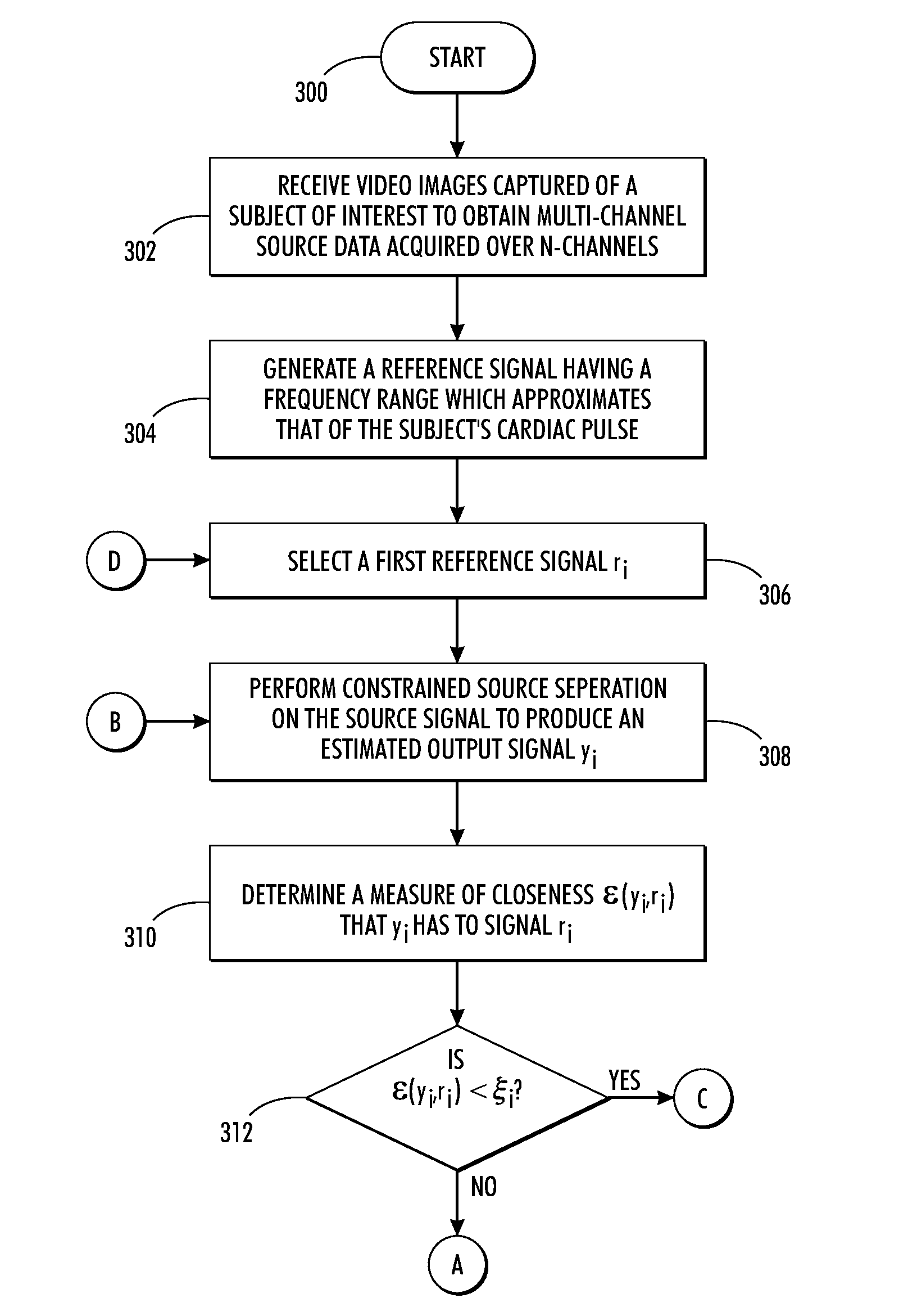
[0068]Reference is now being made to FIG. 10 which illustrates a block diagram of one example processing system 1000 capable of implementing various aspects of the present method described with respect to the flow diagrams of FIGS. 3 and 4.

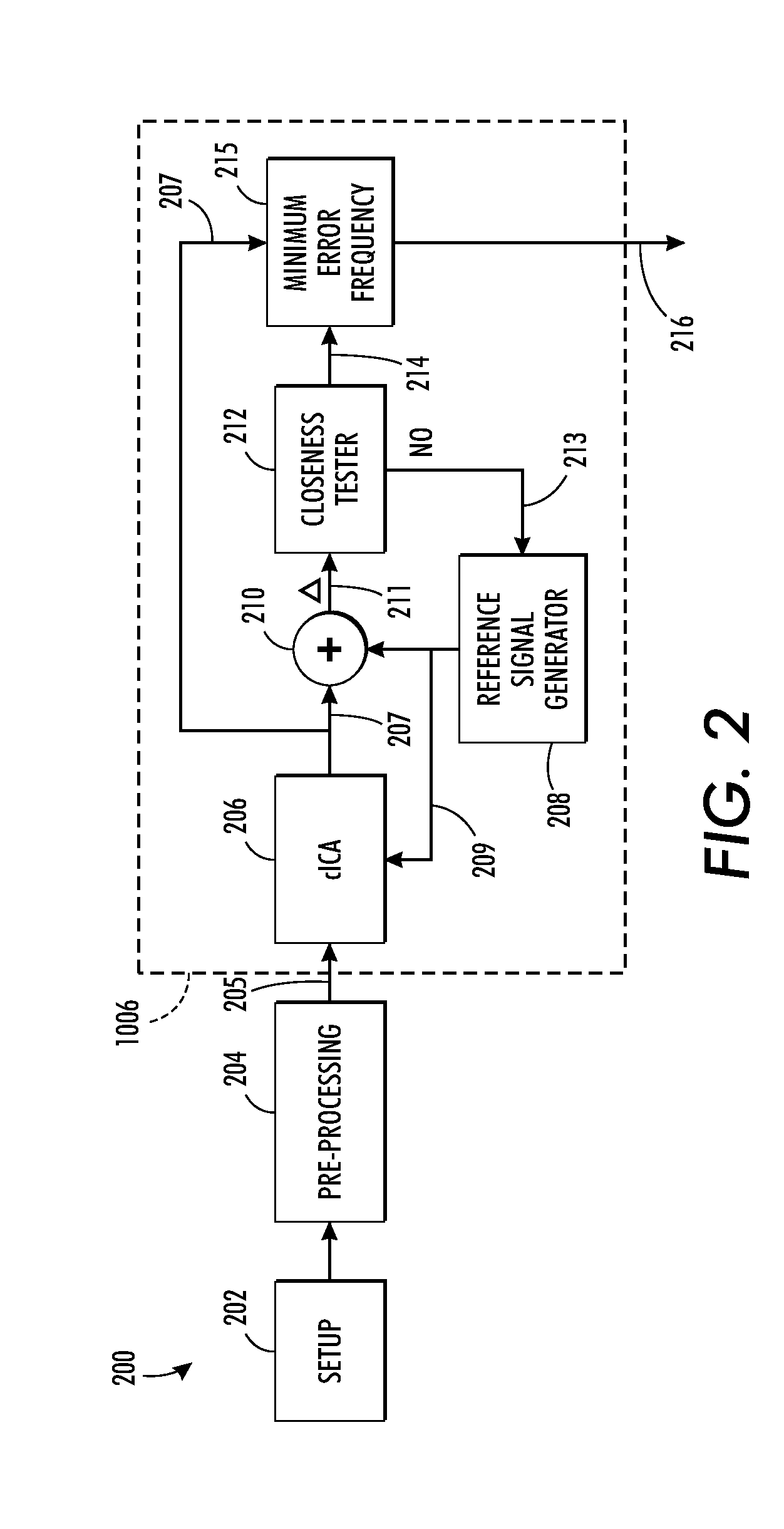
[0069]The embodiment of FIG. 10 receives a sequence of video images captured of a subject of interest intended to be monitored for cardiac function. In FIG. 10, the captured video images are either a plurality of multi-spectral images 1002 captured using a multi-spectral camera or a plurality of RBG images captured using a standard 3-channel camera. The sequence of images collectively comprises a multi-channel source data acquired over a duration of time. Signal processing system 1004 receives the multi-channel source data into source signal recovery module 1006 which performs all the functionality as describe in FIG. 2. Memory 1008 and CPU 1010 facilitate all of the processing and outputs the final estimated cardiac frequency 216. Th...


PUM


Abstract

What is disclosed is a system and method for recovering a patient's cardiac pulse rate from a sequence of video images recording of that patient. In one embodiment, a reference signal of a particular frequency is generated at predetermined frequency intervals and a constrained source separation is performed on the source data to obtain an estimation of the source signal intended to be recovered. The reference signal is updated and constrained source separation is again performed. These operations are repeated for all frequencies of the reference signal. The frequency at which a minimum error is achieved is determined to be the subject's recovered cardiac pulse frequency. In such a manner, the source signal is extracted and recovered reliably from captured multi-channel RGB signals or multispectral signals. The teachings hereof find their uses in a variety of medical solutions including various military, security and telemedicine applications. Various embodiments are disclosed.

Description

TECHNICAL FIELD[0001]The present invention is directed to systems and methods for recovering an estimated cardiac pulse rate from a sequence of RGB or multi-spectral video image data captured from the facial / skin region of a person or animal being monitored for cardiac function in a remote sensing environment.BACKGROUND[0002]Assessment of cardio function is vitally important in monitoring neonatal patients, burn or trauma patients, sleep studies, and other cases where a continuous measurement of the heart rate is required. Currently, cardiac pulse is measured using an electrocardiogram (ECG) which often requires adhesive patches, spring loaded clips, chest straps, and the like, which may prove uncomfortable to patients over the long term. The ability to monitor a patient's physiological signals by non-contact means is highly desirable in the healthcare industry. Although non-contact methods may not be able to provide details concerning cardiac electrical conduction that ECG offers, ...

Claims


Application Information

Patent Timeline
Patent Type & Authority Patents(United States)
IPC IPC(8): A61B5/02
CPCA61B5/0077A61B5/024A61B5/0022A61B5/0033A61B2576/00G01J5/026G16H40/67G01J5/07
Inventor MESTHA, LALIT KESHAVKYAL, SURVITSOURI, GILL RAFAELDIANAT, SOHAIL A.XU, BEILEI
Owner XEROX CORP
Learn More