
Maternal-fetal monitoring system

a monitoring system and mother-fetus technology, applied in the field of mother-fetus monitoring system, can solve the problems of limited predictive value of tests, affecting the health of the fetus, and entanglement of patient expense and inconvenience, so as to reduce the risk of the fetus' health and eliminate uncomfortable equipmen

Active Publication Date: 2005-12-01
UNIV OF FLORIDA RES FOUNDATION INC +1
View PDF38 Cites 194 Cited by
  • Summary
  • Abstract
  • Description
  • Claims
  • Application Information

AI Technical Summary

Benefits of technology

[0021] In a preferred embodiment, the system also incorporates an intelligence means, such as a neural network system, that utilizes the clinical data to analyze and interpret fetal well-being (both antepartum and intrapartum). The intelligence means can also offer clinical advice including, but not limited to, fetal distress, maternal distress, labor progress, contraction effectiveness, uterine rupture, likelihood of delivery within a defined period (i.e., preterm evaluation), and likelihood of successful vaginal delivery. Furthermore, immediate postpartum monitoring of the EHG can provide the user with information regarding uterine tone (i.e., pre-delivery and / or post-delivery). For example, the subject monitoring system can signal uterine atony and prompt early intervention.
[0022] The present invention provides comprehensive monitoring / classification of maternal-fetal health through the use of novel methods for accurately extracting maternal ECG, heart rate and uterine activity (EHG), as well as fetal ECG, heart rate, and positioning, including (1) using an independent component analysis algorithm (ICA) to separate maternal and fetal signals; (2) using signal processing means for automatically determining the fetal and maternal channels, (3) using signal processing means for providing contraction information (4) using signal processing means for providing undistorted ECG waveforms (P and T waves and QRS complex) post-ICA source separation; (5) using a surface electrode mesh to provide accurate positioning and repeatability in extracting fetal ECG signals, higher signal quality and higher separation performance, and enhance patient comfort; and (6) using a neural network to present clinical observations regarding maternal and fetal health (i.e., fetal distress, maternal distress, labor progress, contraction effectiveness, likelihood of delivery within a defined period) in view of clinical services to be provided.
[0023] The algorithms utilized in the present invention are particularly advantageous in that they enable the maternal-fetal monitoring system to provide real-time results as well as automatic and real-time classification of maternal and fetal channels of output using portable, low power digital signal processor implementations.
[0025] The maternal-fetal monitoring system of the present invention is particularly advantageous as either an add-on or stand-alone monitor for use in a variety of settings. As an all-inclusive monitoring system, the maternal-fetal monitoring system of the present invention can be effectively used in place of non-stress tests, contraction stress tests, biophysical profiles, preterm labor evaluations, and existing fetal monitoring systems. Moreover, by providing an all-inclusive monitoring system, which includes an intelligence means for offering clinical advice, the present invention can provide improved prediction of maternal / fetal well-being as well as prediction of labor progress as compared to existing fetal monitoring systems. The system also provides the ability to monitor maternal and / or fetal well-being during surgery (both cesarean section and non-obstetric surgery in the pregnant patient) without interfering with the sterile field. In addition, the system provides the only means to continuously monitor postpartum uterine tone.
[0027] The maternal-fetal monitoring system of the present invention enables non-invasive capture of fetal ECG results with much less risk to the fetus' health and no limitation on cervical dilation or ruptured membranes, as is generally presented with use of current systems and methods for monitoring fetal ECG. The system eliminates uncomfortable equipment, and can be used intraoperatively during Cesarean delivery as well as during non-obstetric surgery in the pregnant patient (i.e., appendectomy or cholecystectomy where ultrasound would be in the sterile field).

Problems solved by technology

While most patients will deliver a healthy child with or without monitoring, more than 5 out of every 1,000 deliveries of a viable fetus near term is stillborn, with half having an undetermined cause of death.
Despite their wide acceptance, these tests offer limited predictive value, and give only a glimpse of the fetus at the time of testing.
For high risk patients, once or twice weekly surveillance is often indicated, entailing both expense and inconvenience for the patient.
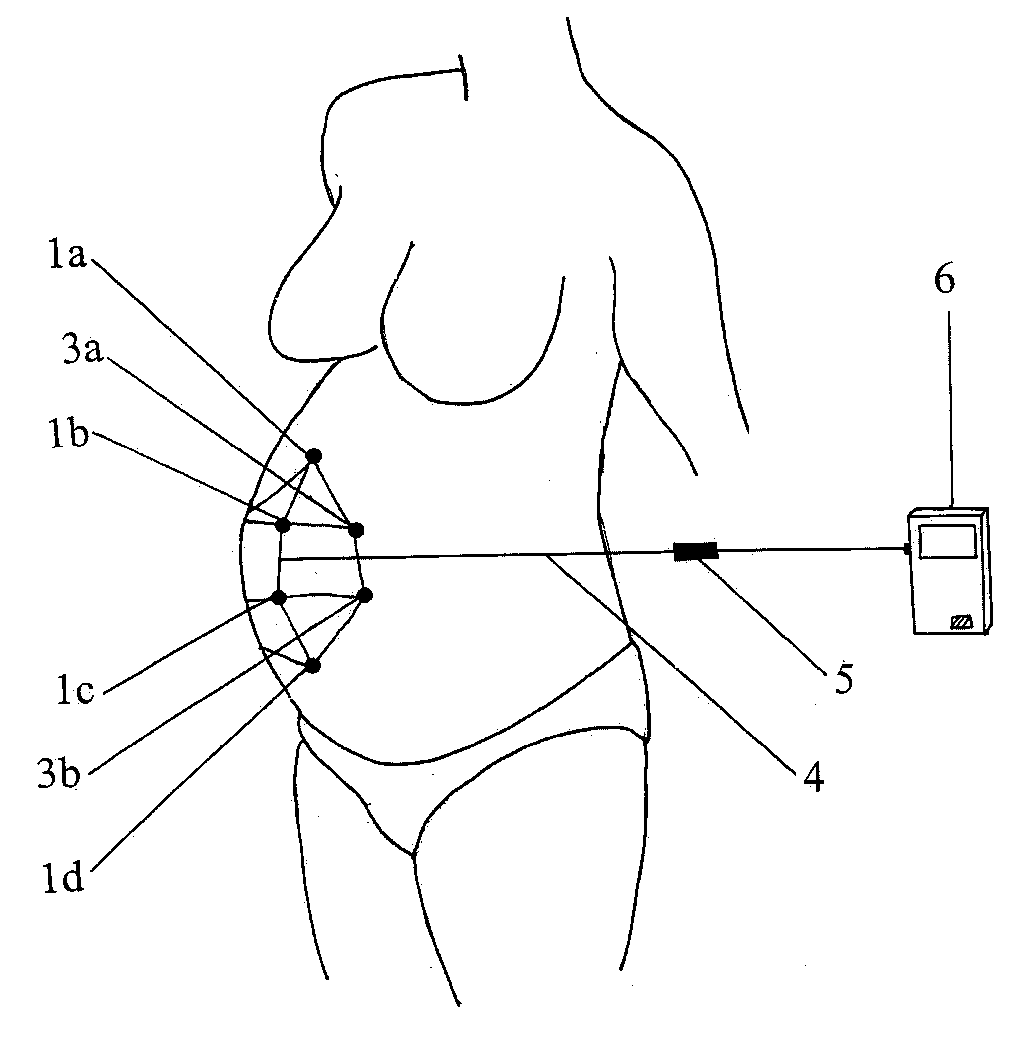
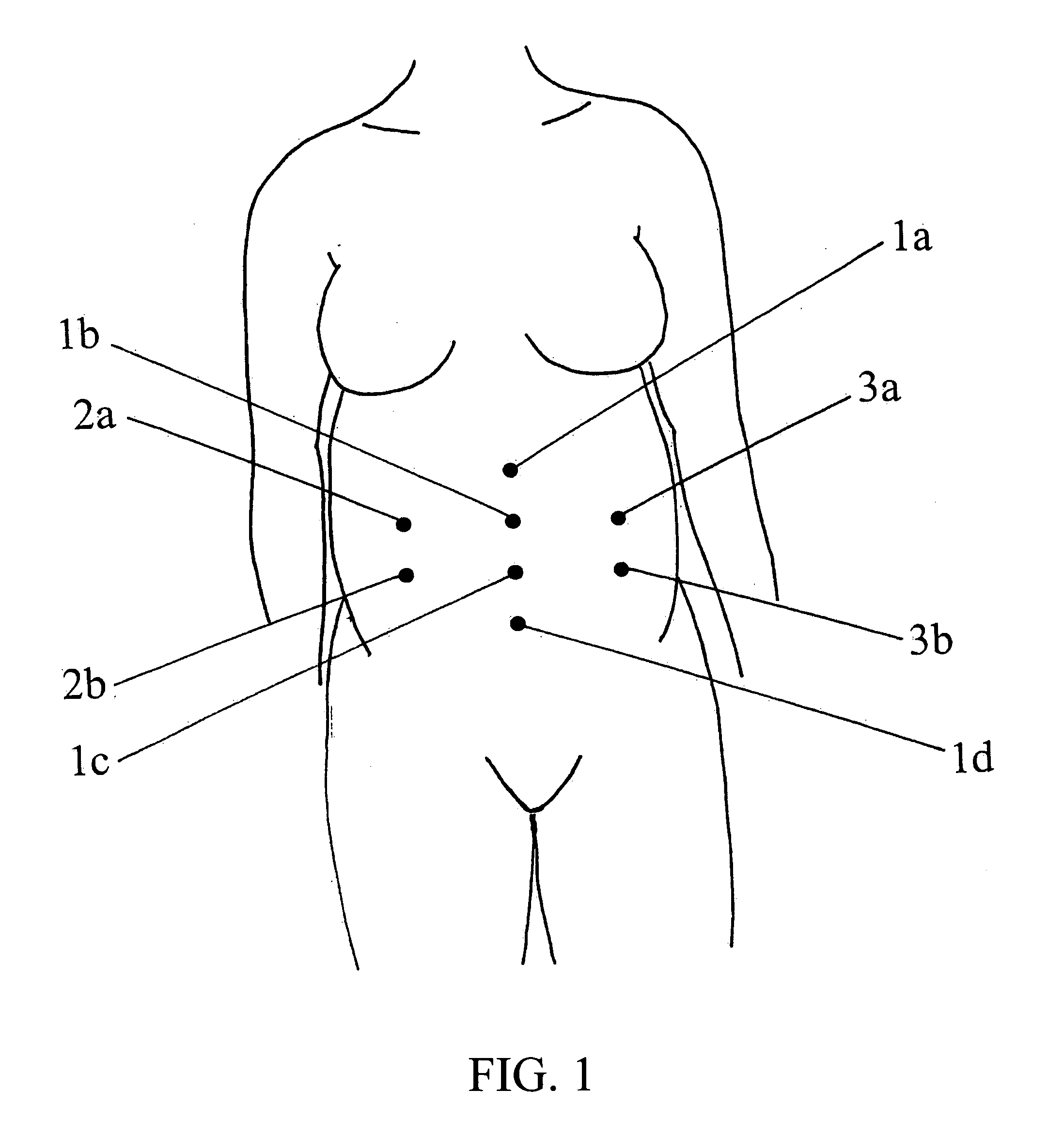
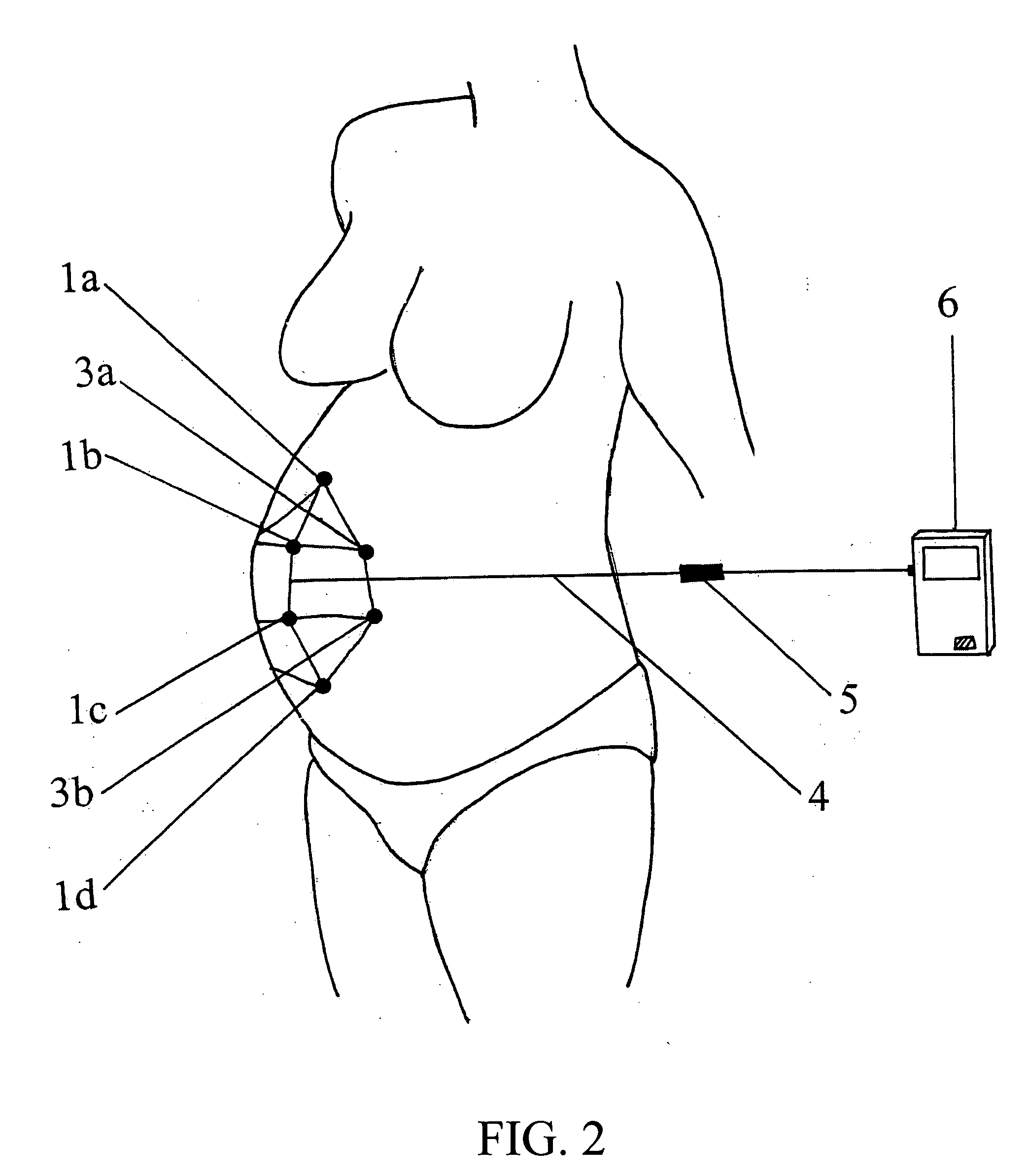
When indicated, more invasive monitors are available, but require ruptured membranes / adequate cervical dilation, and entail some risk, primarily infectious.
The tocodynamometer detects only the presence or absence of tension on the abdomen (whether from uterine contraction or maternal movement), and often fails in the presence of obesity.
Unfortunately, numerous studies have failed to identify this improved outcome with the use of EFM in low-risk deliveries.
Unfortunately, acquisition of the FECG was through the fetal scalp electrode described above which is both invasive and limited in its application.
Non-invasive acquisition of the FECG is a recognized issue of mixed signals.
These methods, unfortunately, do not reliably enable continuous extraction of maternal-fetal data or cannot capture a comprehensive account of maternal-fetal health based on a combination of test results (i.e., fetal heart rate, fetal ECG, maternal ECG, and maternal uterine activity (EHG)).
Unfortunately, magnetocardiography is limited in application, technologically complex, and difficult to administer to assess accurate fetal ECG readings.

Method used


Image

  • Maternal-fetal monitoring system
  • Maternal-fetal monitoring system
  • Maternal-fetal monitoring system

Examples


example 1

Method for Personalizing a Sensor Mesh of the Invention

[0180] In one embodiment of the invention, a mesh of 20 electrodes was used to collect ECG signals. Each of the electrodes was selectively multiplexed into / out of the ICA and Pan Tompkins algorithm operations, depending on the quality of the signal and the TF for the fetal ECG output from Pan Tompkins operator(s). At the output of the ICA algorithm operator(s), the fetal ECG signal was correlated with 8 prefiltered raw signals. The 8 correlation coefficients correspond to the influence of the fetal heart in the different electrodes. The electrodes with the lowest correlation coefficients were switched out of the ICA / Pan Tompkins algorithm operations and other electrodes were switched into those operations to find the best set of electrodes for finding the fetal ECG.

[0181] By providing a method for personalizing a sensor mesh, the subject invention combines cumbersome individual sensor leads into a single cable, to enable the s...

example 2

Maternal-Fetal Monitoring System of the Invention

[0182] As illustrated in FIG. 14, a maternal-fetal monitoring system 200 of the present invention is provided, wherein the system comprises an electrode mesh 202, an amplifier, at least one filter 204, and a processor for running an ICA algorithm 206, preferably Mermaid, and calculating a Trust Factor 208, to provide clinical information regarding maternal-fetal health. The electrode mesh has 10 electrodes, which are placed on the maternal abdomen. Electrodes having offset tabs that aid in eliminating noise artifacts are preferred. In this example, 1″¼ Blue sensor Ag / AgCl electrodes are used. A 10 lead shielded cable is used to connect the electrodes to the amplifier.

[0183] The amplifier of the subject invention takes into account the fetal ECG, which typically is around 10 μV to 50 μV in amplitude. In a preferred embodiment, the amplifier of the invention is a high-resolution, low-noise unipolar amplifier in which all channels are ...

example 3

Comparison of Intra-Uterine Pressure Values Using a Neural Network of the Subject Invention versus an Intra-Uterine Pressure Catheter (IUPC)

[0195] An intelligence system (i.e., neural network system) has been trained to predict the IUP from the electrical signals obtained from the abdominal ECG electrodes (EHG). The network was trained with three different patients and can be used to create an estimated non-invasive measure of IUP in patients. FIGS. 15A-C show the output of the neural network and the IUPC signal for three different data sets A, B, and C. The mean squared error (MSE) and correlation coefficient (CorrCoef) for each data set are provided in Table 1. As can be seen most of the peaks in a commercially available IUP catheter signal are well captured by the neural network of the subject invention. The high correlation coefficient in test data further supports the fact that the network has learnt the relationship.

TABLE 1Quantitative Comparison of Values from an MLP of th...


PUM


Abstract

A maternal-fetal monitoring system for use during all stages of pregnancy, including antepartum and intrapartum stages. The maternal-fetal monitoring system of the subject invention comprises (1) a set of sensors; (2) an amplifying / filtering means; (3) a computing means; and (4) a graphical user interface. Accurate clinical data, which can be extracted and provided to the user in real-time using the system of the invention, include without limitation, maternal electrocardiogram (ECG) signals, maternal uterine activity signals (EHG), maternal heart rate, fetal ECG signals, and fetal heart rate. In a preferred embodiment, the maternal-fetal monitoring system of the invention includes an intelligence means, such as a neural network system, to analyze and interpret clinical data for use in clinical diagnosis antepartum, intrapartum and postpartum, as well as delivery strategy.

Description

GOVERNMENT SUPPORT [0001] This invention was made with government support under a grant awarded from the National Science Foundation under grant number 023960. The government has certain rights in the invention.BACKGROUND OF INVENTION [0002] Assessment of the fetus during pregnancy, and particularly during labor and delivery, is an essential but yet elusive goal. While most patients will deliver a healthy child with or without monitoring, more than 5 out of every 1,000 deliveries of a viable fetus near term is stillborn, with half having an undetermined cause of death. (National Vital Statistics System (NVSS), CDC, NCHS as published in “Healthy People 2010, Understanding and Improving Health: Chapter 16,” co-authored by the Centers for Disease Control and Prevention and Health Resources and Services Administration, 2nd Edition, U.S. Government Printing Office, November 2000). The risk of this unfortunate consequence is increased in a subgroup of “high risk” patients (e.g. diabetics)...

Claims


Application Information

Patent Timeline
IPC IPC(8): A61B5/344A61B5/00A61B5/024A61B5/03A61B5/103A61B5/117A61B10/00G06F17/00
CPCA61B5/0011A61B5/02411A61B5/033A61B5/0444A61B5/7264A61B5/04882A61B5/4362A61B5/6805A61B5/6831A61B5/0452A61B5/344A61B5/349A61B5/391
Inventor MAROSSERO, DOROTHEEEULIANO, TAMMY Y.EULIANO, NEIL RUSSELL IIPRINCIPE, JOSE C.
Owner UNIV OF FLORIDA RES FOUNDATION INC
Learn More