Serial multiple antigen colocalization in paraffin-embedded tissue

a paraffin-embedded tissue and antigen colocalization technology, applied in the field of paraffin-embedded tissue antigen colocalization, can solve the problems of limited, negative effects, and limitations of current colocalization methods, and achieve the effect of less tissue damag

Active Publication Date: 2014-06-17
UNIV OF VIRGINIA ALUMNI PATENTS FOUND
View PDF3 Cites 16 Cited by
  • Summary
  • Abstract
  • Description
  • Claims
  • Application Information

AI Technical Summary

Problems solved by technology

Current colocalization methods, however, have several limitations.
The most commonly used method, multi-color immunofluorescence (MCIF), is limited by the number of viable combinations of available fluorescent tags and can be negatively affected by spectral bleed-through, antibody cross-reactivity, photo-bleaching, and autofluorescence of paraffin-embedded tissue (3).
Because of these limitations, this method is usually restricted to the simultaneous visualization of just two antigens plus a nuclear counterstain.
Moreover, multiple chromogenic substrates do not allow colocalization in the same cellular compartment due to the obscuring of lighter colors by darker chromogens.
Additionally, the use of more than one primary antibody from the same species (i.e., mouse or rabbit) is usually impossible due to secondary antibody cross-reactivity.

Method used


Image

  • Serial multiple antigen colocalization in paraffin-embedded tissue
  • Serial multiple antigen colocalization in paraffin-embedded tissue
  • Serial multiple antigen colocalization in paraffin-embedded tissue

Examples


embodiments

[0129]The ability to simultaneously visualize expression of multiple antigens in cells and tissues can provide powerful insights into cellular and organismal biology. However, standard methods are limited to the use of just two or three simultaneous probes and have not been widely adopted for routine use in paraffin-embedded tissue.

[0130]The present invention allows for analyzing multiple targets in a reasonable amount of time, but because there is a wide range in the timing of antibody incubations and washes, the number of antigens being targeted, etc., the amount of total time required for the whole process can vary. The present invention usually requires about 1-2 hours per round, resulting in 5-10 hours for a 5 antigen experiment. There is also a range of time for scanning each stain, depending on speed of scanner, etc.

[0131]The present invention as described below, wherein a large area is scanned on the exemplified tissue sample on a slide, can be adapted to allow multiples of ...

examples

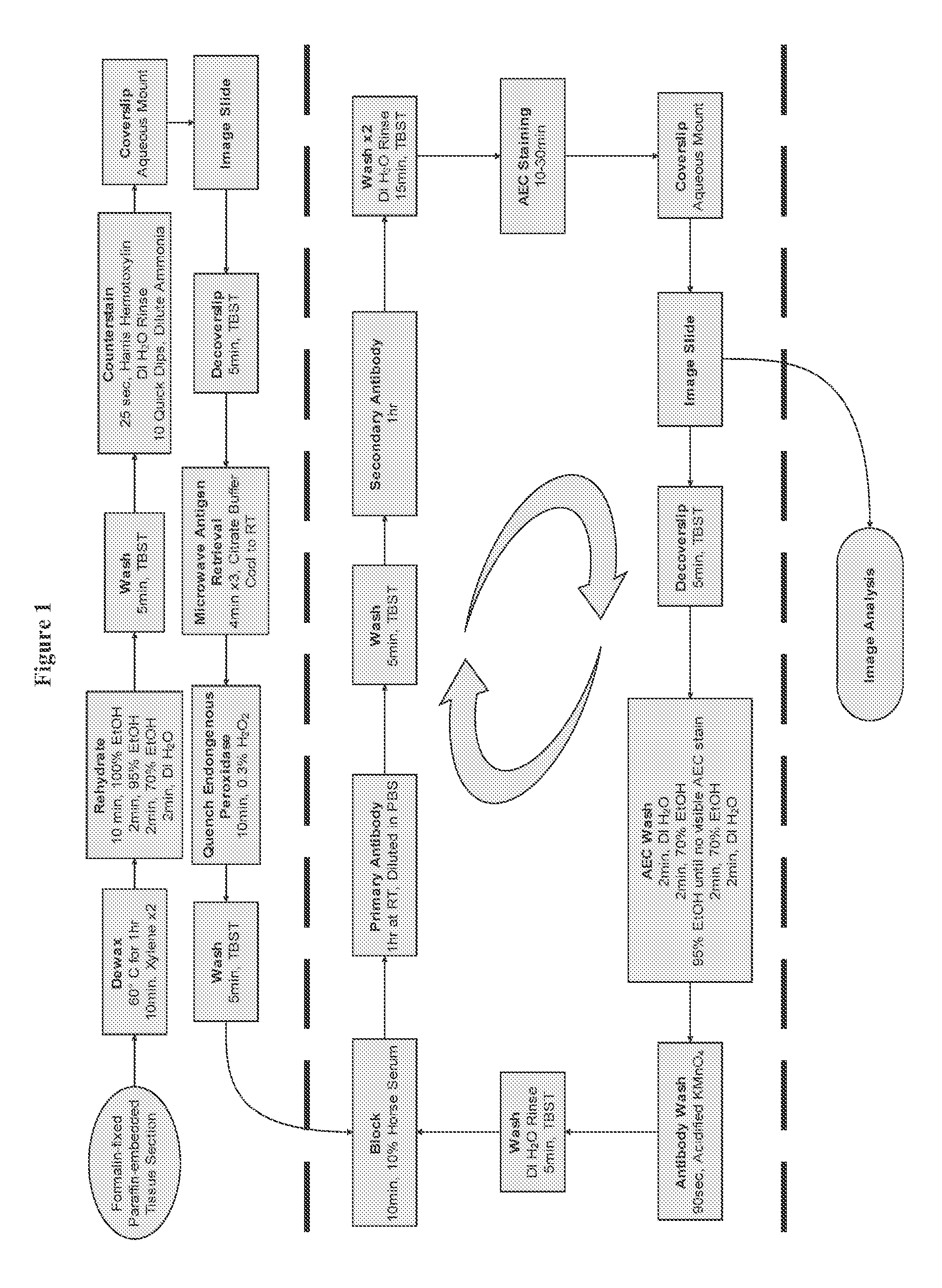
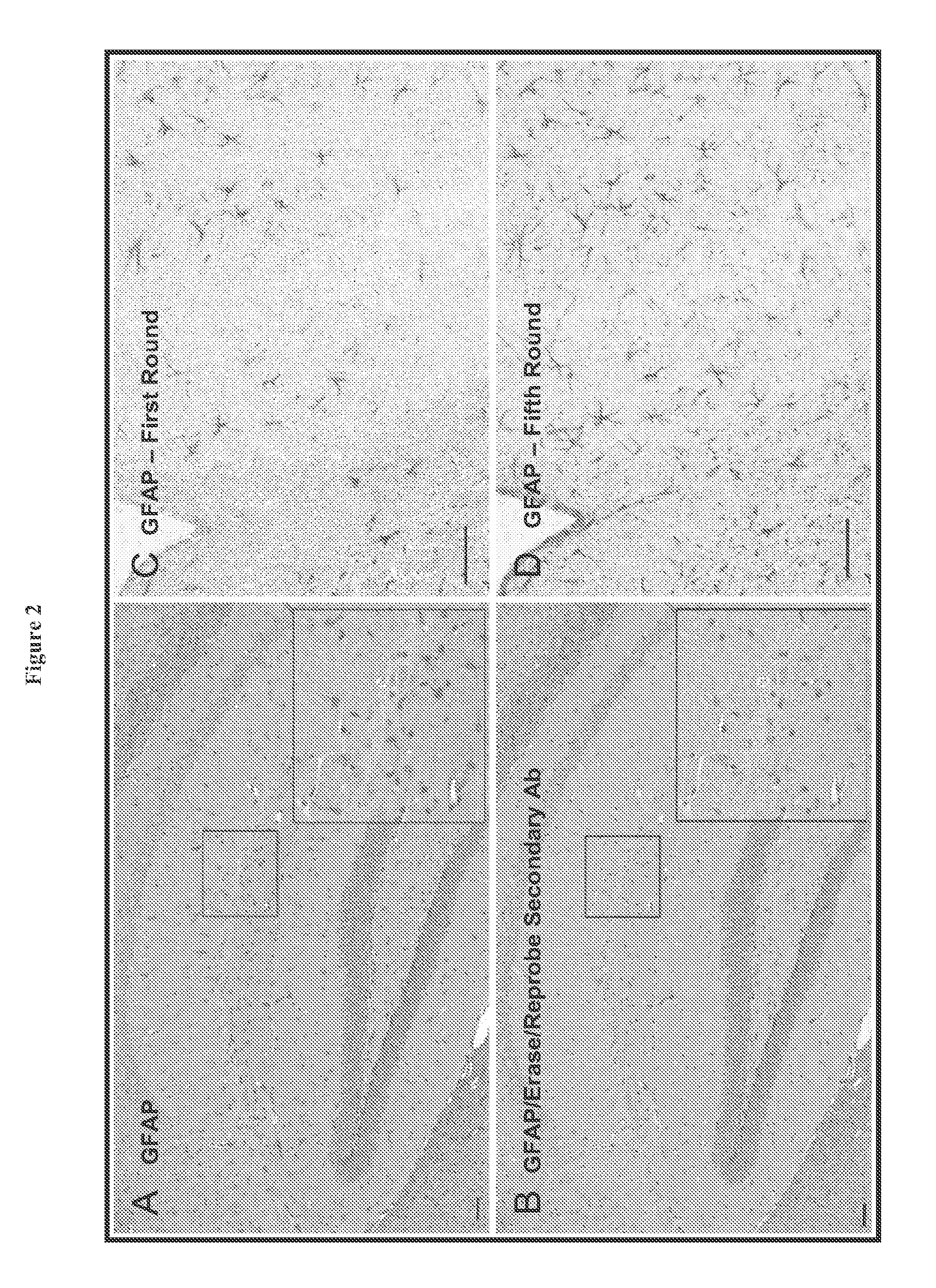
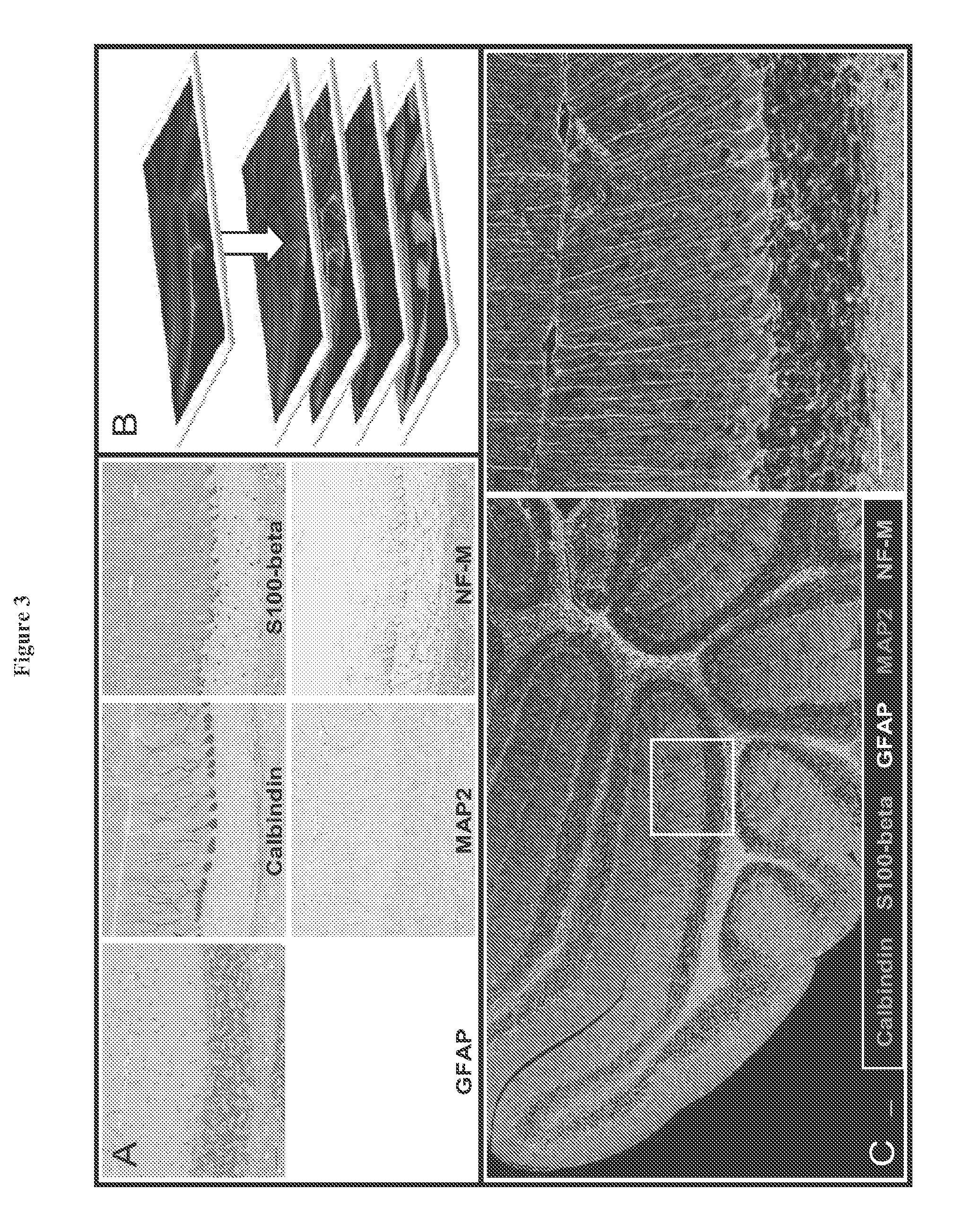
[0184]In order to overcome the limitations described in the Background, described herein is a novel approach called Sequential IMmunoPeroxidase Labeling and Erasing (SIMPLE) that enables the simultaneous visualization of multiple markers within a single tissue section. By combining the use of an alcohol-soluble immunoperoxidase substrate, 3-amino-9-ethylcarbazole (AEC), with a previously described antibody-antigen dissociation method (6), we have performed up to five serial rounds of staining on a single tissue section without loss of tissue antigenicity. This method is accessible to any histology lab and requires no specialized equipment. The use of a whole slide digital scanning microscope greatly increases the power of SIMPLE as it facilitates permanent archiving of each round of labeling. In addition, from a practical standpoint, the method enables multiple immunohistochemical analyses to be performed on otherwise limited tissue samples, such as very small biopsies and tissue mi...


PUM

PropertyMeasurementUnit
pHaaaaaaaaaa
pHaaaaaaaaaa
solubleaaaaaaaaaa

Abstract

The present invention provides a novel method called Sequential IMmunoPeroxidase Labeling and Erasing (SIMPLE) that enables the simultaneous visualization of at least five markers within a single tissue section. Utilizing the alcohol-soluble peroxidase substrate 3-amino-9-ethylcarbazole (AEC), combined with a rapid non-destructive method for antibody-antigen dissociation, the present application discloses the ability to erase the results of a single immunohistochemical stain while preserving tissue antigenicity for repeated rounds of labeling. The present invention also provides methods for visualizing multiple antigens simultaneously.

Description

CROSS REFERENCE TO RELATED APPLICATIONS[0001]This application is a national stage filing of International Application No. PCT / US2010 / 029763, filed Apr. 2, 2010, which is entitled to priority pursuant to 35 U.S.C. §119(e) to U.S. provisional patent application No. 61 / 166,384, filed on Apr. 3, 2009. The entire disclosures of the afore-mentioned patent application are incorporated herein by reference.BACKGROUND[0002]Visual colocalization of molecular species within sectioned tissue can provide insights into cellular biochemistry and can serve as the basis for further study of protein-organelle and protein-protein interactions. Visualization of abnormal protein coexpression in neoplastic tissue may elucidate components of oncogenic signaling pathways. Colocalization of COX2 and laminin-5, for example, has been observed at the cancer-stromal interface of lung adenocarcinoma and may be associated with abnormalities in p53 expression (1). Additionally, colocalization of β-catenin with comp...

Claims


Application Information

Patent Timeline
Patent Type & Authority Patents(United States)
IPC IPC(8): G01N33/50G01N33/53G01N33/58
CPCG01N33/581G01N33/5023G01N2800/60
Inventor PAPIN, JASON A.MANDELL, JAMES W.GLASS, GEORGE F.
Owner UNIV OF VIRGINIA ALUMNI PATENTS FOUND
Why Eureka
Try Eureka