System and method reconstructing a nuclear medicine image using deformed attenuation image

a nuclear medicine and attenuation image technology, applied in the field of tomographic imaging, can solve the problems of increasing image noise, artifacts and/or contrast dilution of lesions from motion blurring, and not generally providing structural details of pet imaging as well as other types of scanners, and achieve the effect of improving quantitative accuracy in tomographic imaging

Active Publication Date: 2015-05-12
KINGS COLLEGE LONDON +1
View PDF18 Cites 8 Cited by
  • Summary
  • Abstract
  • Description
  • Claims
  • Application Information

AI Technical Summary

Problems solved by technology

However, PET imaging does not generally provide structural details as well as other types of scanners such as CT and MRI scanners.
Respiratory motion may result in artifacts and / or contrast dilution of lesions from motion blurring.
However, the reduction in blur may come at the expense of increased image noise because each gate has fewer counts than the un-gated image.
Although the RRA approach using respiratory gated PET data may address respiratory motion blur, a respiratory phase mismatch may exist between an attenuation image that is used in a PET image reconstruction process and each respiratory gated PET data set since the attenuation image may be obtained from a CT image, which is usually taken while the subject is holding his or her breath.
This mismatch may generate undesirable artifacts in reconstructed PET images for each gate or the output PET image of the RRA approach, preventing accurate PET quantitation.

Method used


Image

  • System and method reconstructing a nuclear medicine image using deformed attenuation image
  • System and method reconstructing a nuclear medicine image using deformed attenuation image
  • System and method reconstructing a nuclear medicine image using deformed attenuation image

Examples


Embodiment Construction

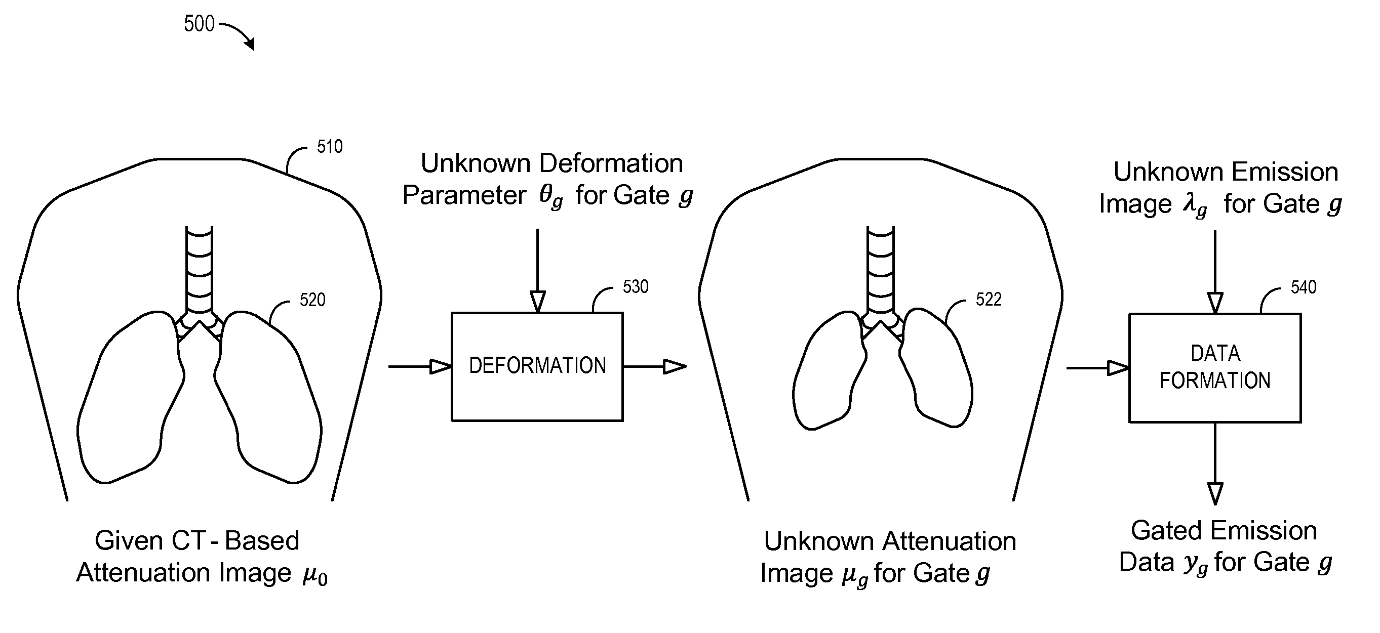
[0018]Embodiments disclosed herein include an imaging method for providing motion correction in tomographic imaging. Some embodiments are associated with reconstruction of respiratory-gated PET images using a CT image for PET attenuation correction. Note that patient motion may lead to a mismatch between PET and CT images and result in artifacts in reconstructed PET images. This mismatch may, according to some embodiments, be adjusted between gated PET and CT images. The mismatch adjustment may be performed, for example, in the CT-based attenuation image to correct for motion in a region of interest. An attenuation mismatch may be modeled as a deformation of the initially given CT-based attenuation image and the deformation as well as the PET image for each gate may be jointly estimated from respiratory gated PET data. The resulting estimated deformation may yield a mismatch corrected attenuation image, which may result in a PET image free of attenuation mismatch induced artifacts. ...


PUM


Abstract

According to some embodiments, an emission tomography scanner may acquire emission scan data. One or more anatomical images may be generated using an anatomical imaging system, and the anatomical images may be processed to obtain an initial attenuation image. An emission image and a corrected attenuation image may be jointly reconstructed from the acquired emission scan data, the corrected attenuation image representing a deformation of the initial attenuation image. A final reconstructed emission image may then be calculated based on the reconstructed emission image and / or the corrected attenuation image. The final reconstructed emission image may then be stored in a data storage system and / or displayed on a display system.

Description

BACKGROUND[0001]The invention relates generally to tomographic imaging and, more particularly, to methods and systems for correcting for motion-induced attenuation mismatch for use in tomographic imaging.[0002]Tomographic imaging has become an integral part of healthcare services. Examples of tomographic imaging include Positron Emission Tomography (“PET”) imaging, Single Photon Emission Computed Tomography (“SPECT”) imaging, X-ray Computed Tomography (“CT”) imaging, and magnetic resonance imaging (“MRI”). In CT imaging, X-rays are propagated through the body and are detected on the other side of the body. The X-rays are attenuated to different degrees depending on encountered bodily structures, resulting in an image showing the structural features of the body. MRI also creates images of the internal structures of the human body by exploiting the property of nuclear magnetic resonance. MRI provides good contrast resolution by using various optimized radio frequency pulse sequences. ...

Claims


Application Information

Patent Timeline
Patent Type & Authority Patents(United States)
IPC IPC(8): G06K9/00G06T11/00
CPCG06T11/003G06T11/005
Inventor MANJESHWAR, RAVINDRA MOHANAHN, SANGTAETHIELEMANS, KRIS
Owner KINGS COLLEGE LONDON
Why Eureka
Try Eureka