
Bladder cancer tumor marker, antibody and use thereof

a tumor marker and bladder cancer technology, applied in the field of tumor immunology, can solve the problems of inability to achieve the overall therapeutic effect, inconvenient treatment process, toxic and side effects, etc., and achieve the effect of inhibiting the growth facilitating malignant transformation of bladder cancer, and effectively inhibiting the proliferation of the bladder cancer cell line t24

Active Publication Date: 2015-07-07
INSITUTE OF BIOPHYSICS CHINESE ACADEMY OF SCIENCES
View PDF10 Cites 0 Cited by
  • Summary
  • Abstract
  • Description
  • Claims
  • Application Information

AI Technical Summary

Benefits of technology

The invention describes a new antibody called BCMab1 that can treat bladder cancer by inhibiting its growth. The antibody was tested on in-vitro cultured T24 cells and a nude mouse tumor model, and was found to be effective in preventing the cancer's propagation. The patent also discusses a key protein called glycosyltransferase GALNT1 that is highly expressed in bladder cancer and its role in promoting malignant transformation. The patent proposes that targeting GALNT1 could be a potential therapeutic strategy for bladder cancer. Overall, the invention offers a new tool for diagnosis, prognosis, and immunotherapy of bladder cancer.

Problems solved by technology

Moreover, all these treating processes have apparent toxic and side-effects.
Although the above classes of medicaments, used either alone or in combination, achieved some effects in the reduction of the relapse rate after the operation of bladder cancer, the overall therapeutical effect is not desirable due to the presence of many issues such as low specificity, multiple drug resistance (MDR) of tumors, and so on.
Moreover, because of their low molecular weights, the above classes of medicaments not only can act on the entire bladder and urethra unspecifically, but also can be absorbed by the mucous membranes of bladder and urethra, which easily results in topical or systematic toxic and side-effects.
For the past several years, the molecular markers, such as nuclear matrix protein, bladder tumor antigen and satellite instability etc. newly involved in clinic were also limited by their low sensitivities and specificities.

Method used


Image

  • Bladder cancer tumor marker, antibody and use thereof
  • Bladder cancer tumor marker, antibody and use thereof
  • Bladder cancer tumor marker, antibody and use thereof

Examples


example 1

Preparation and Purification of BCMab1 Monoclonal Antibody

(1) Preparation of Hybridoma

[0074]Balb / C mice (commercially purchased from Vital River Laboratory Animal Technology Co. Ltd., Beijing) were immunized with human bladder cancer cell line T24 (ATCC: HTB-4) by intraperitoneally injecting a dose of 1×10⁷ cells / ml PBS. After two weeks, the mice were immunized again with the same injection volume and method. While the serum titer of the mice met the requirement, cell fusion was arranged. The mice were boosted three days before the fusion. At the same time, mouse myeloma cells Sp2 / 0 (ATCC CRL-1772) were prepared.

[0075]The sensitized B lymphocytes were fused with the myeloma cells (Practical Immunology, Yang, Y. ed., Changchun Press, published in December, 1994), and selectively cultured (using peritoneal macrophages of the mice as feeder cells) in HAT medium (purchased from Invitrogen Co.; HAT is an enumerate of the initials of hypoxantin, aminopterin and thymidin; HAT medium is thu...

example 2

Identification of Monoclonal Antibody BCMab1

[0080]Normal bladder tissue cells HCV29 were purchased from Peking University Health Science Center. Cell lines T24, EJ, HepG2, LoVo, 293, A375, Jurkat, PC-1, MCF-7, K562, Hela, and other cell lines were purchased from ATCC (Rockville, Md., US). Bladder cancer tissues and normal tissues of human were received from The Second Affiliated Hospital of Kunming Medical University, with informed consents. BALB / C mice and nude mice were both purchased from Chinese Academy of Medical Science Institute of laboratory animal.

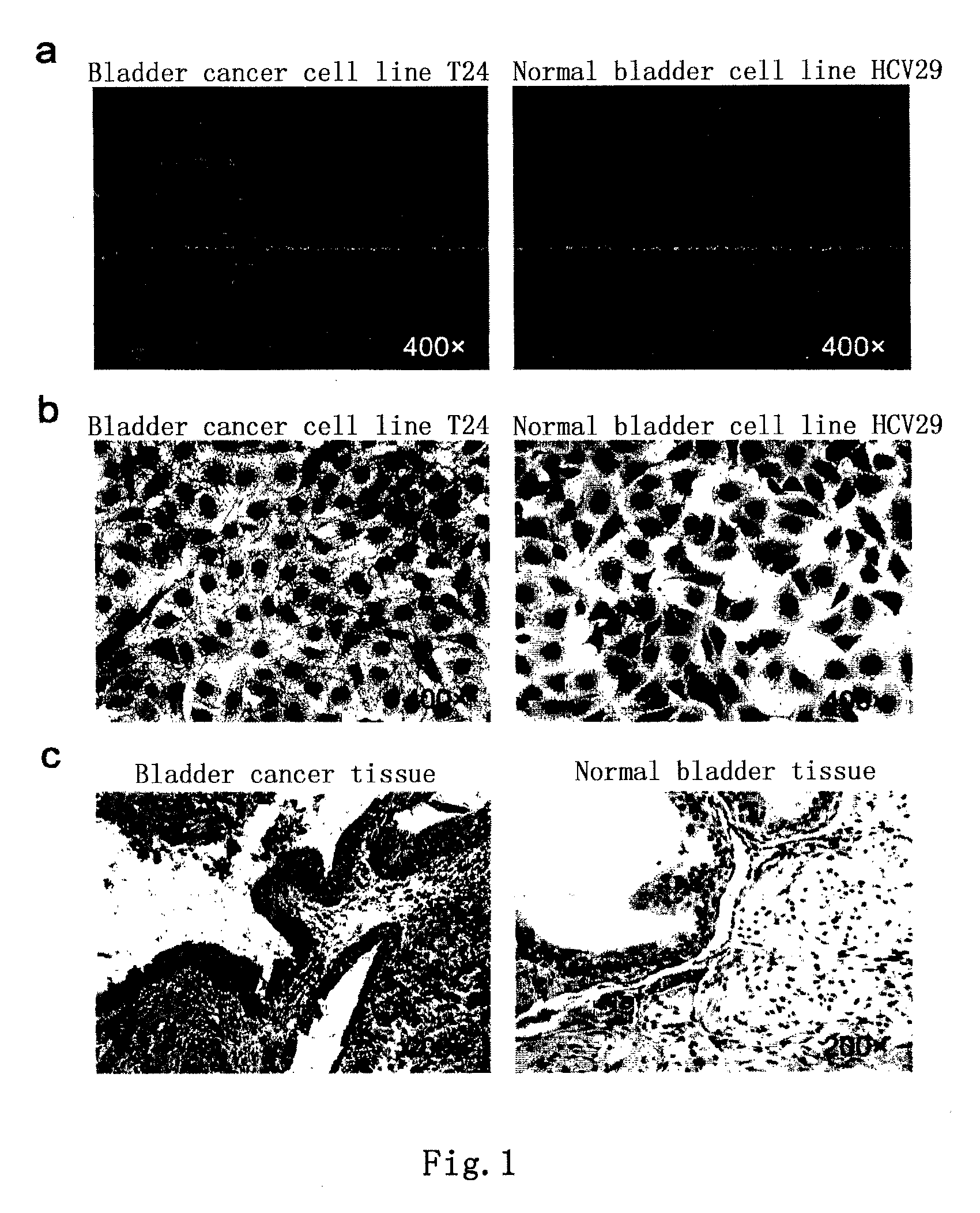
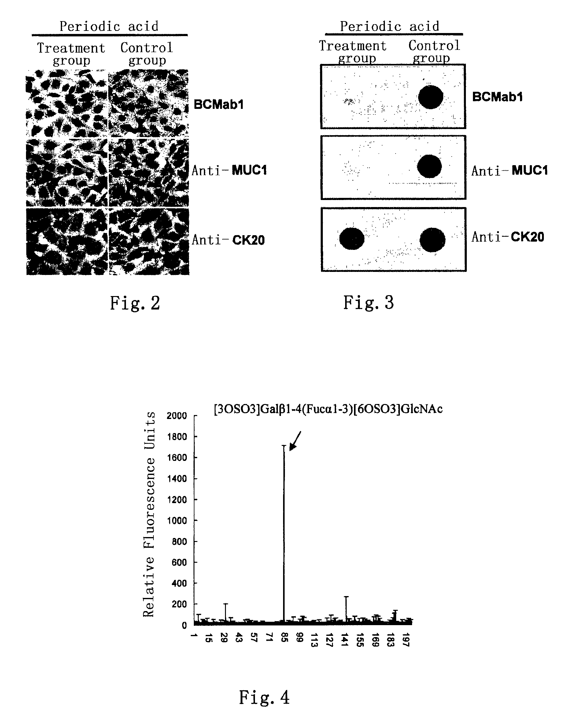
[0081]Bladder cancer tissue slices of human (received from Peking University Third Hospital) were stained immunohistochemically with the monoclonal antibody BCMab1 prepared in Example 1 using a conventional method. The result, as shown in FIG. 1, indicated that the bladder cancer tissues of human showed positive reactivity after stained with the monoclonal antibody BCMab1, a secondary antibody (anti-mouse IgG-HRP) and DAB substrat...

example 3

Identification and Preparation of Antigen AG-α3β1

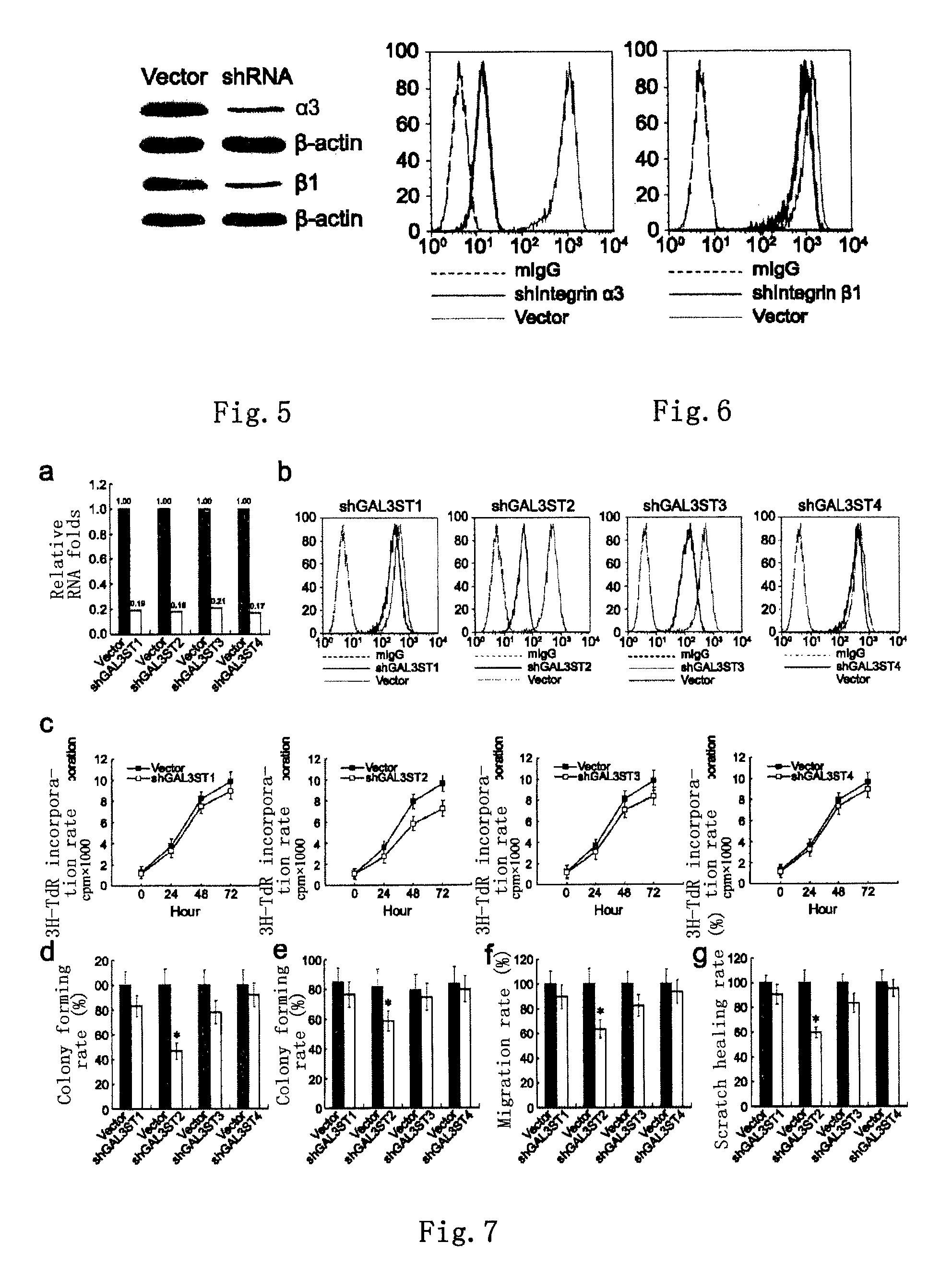
[0084]Human bladder cancer cell line T24 was cultured in a RPMI-1640 medium containing 10% fetal bovine serum (purchased from Invitrogen Co.). After digested with 0.25% pancreatin, 1×10⁸ T24 cells were washed with PBS for 3 times. Then 100 μg monoclonal antibody BCMab1 was added, incubated at 4° C. for 2 hours, and washed with PBS for 3 times. The cells were lysed with 1 ml detergent lysis buffer (50 mM Tris-HCl, pH 8.0; 150 mM NaCl; 0.02% sodium azide; 0.1% SDS; 100 μg / ml PMSF; 1 μg / ml Aprotinin; 1% NP-40; 0.5% sodium deoxycholate) for 30 min, and centrifuged at 12,000 g for 10 min. The supernatant was separated and passed through the Protein G affinity chromatographic column. The column was washed with PBS till its OD value approached zero. Subsequently, elution was carried out using a 0.2 M glycine-HCl solution (pH 2.8), and the eluent was collected. The OD value of each collecting tube was measured, and then the eluent with an OD ...


PUM

PropertyMeasurementUnit
diameteraaaaaaaaaa
body weightaaaaaaaaaa

Abstract

Provided in the present invention are an aberrantly glycosylated integrin, AG-α3β1, and use thereof as a bladder cancer marker. Also provided in the present invention are a hybridoma cell generating an anti-AG-α3β1 monoclonal antibody, a monoclonal antibody BCMab1 secreted by the same, and use of BCMab1 in the preparation of a medicament for the treatment of bladder cancer. Also provided in the present invention is use of inhibitors of GAL3ST2 and N-acetylgalactosaminyltransferase 1 in the preparation of a medicament for the treatment of bladder cancer.

Description

CROSS REFERENCE TO RELATED APPLICATION[0001]This is a national stage application of PCT / CN2011 / 001345, filed on Aug. 12, 2011, which claims priority of Chinese Patent Application No. CN 201010251384.6, filed on Aug. 11, 2010. This application claims the benefits and priority of these prior applications and incorporates the disclosures of these prior applications by reference in their entirety.FIELD OF THE ART[0002]The present invention belongs to a field of tumor immunology. Particularly, the present invention relates to a novel antigen AG-α3β1 and the preparation thereof (AG: Aberrant Glycosylation), and an anti-AG-α3β1 monoclonal antibody BCMab1. It is demonstrated at cytology and histology levels that AG-α3β1 is expressed on the membranes of human bladder tumor cells only, the antibody BCMab1 not only specifically recognizes AG-α3β1, but also effectively inhibits the proliferation of human bladder tumor cells at cellular level in vitro and in an animal model in vivo. The present ...

Claims


Application Information

Patent Timeline
Patent Type & Authority Patents(United States)
IPC IPC(8): G01N33/53C07K16/00C07K14/705C07K16/28C12N9/10C07K16/30A61K39/395A61K45/06A61K51/10A61K47/48C12Q1/02C12Q1/48C12N15/113G01N33/68G01N33/574
CPCC07K16/2842C07K14/7055G01N33/57488G01N33/6854C12N15/1137C12Q1/48C12Q1/025A61K47/48607A61K51/1093A61K45/06A61K39/39558C07K16/3038G01N33/57484C07K14/70546C12N9/1051C12Y204/01041G01N2333/70546G01N2440/38A61K47/6861A61P13/10A61P35/00
Inventor LI, CHONGFAN, ZUSENZHANG, HONGLIANDAI, ZHONGHUATANG, HAIDONGCHEN, JUN
Owner INSITUTE OF BIOPHYSICS CHINESE ACADEMY OF SCIENCES
Learn More