
Internal focus reference beads for imaging cytometry

a technology of imaging cytometry and focus reference beads, which is applied in the field of analytical and monitoring systems, can solve problems such as the inability of imaging systems, and achieve the effects of improving detection limits and confidence, more accurate detection and measurement, and effective and efficient internal focus

Active Publication Date: 2015-07-07
NEXCELOM BIOSCIENCE LLC
View PDF9 Cites 3 Cited by
  • Summary
  • Abstract
  • Description
  • Claims
  • Application Information

AI Technical Summary

Benefits of technology

The invention is a new way to calibrate and focus on cells in images. It improves the detection and measurement of microscopic objects like cells, even at very low concentrations. This system enables effective and efficient internal focus and referencing for confirmation of low concentration leukoreduced blood sample cell count. As a result, the detection limits and confidence on the results obtained from imaging cytometry are significantly improved.

Problems solved by technology

However, most of the leukoreduced patient blood samples contain very low concentration of leukocytes; therefore, an imaging system may not be able to detect any presence of leukocytes, which means there is no object to focus on in the image field.

Method used


Image

  • Internal focus reference beads for imaging cytometry
  • Internal focus reference beads for imaging cytometry
  • Internal focus reference beads for imaging cytometry

Examples


example 1

Imaging of Leukocytes

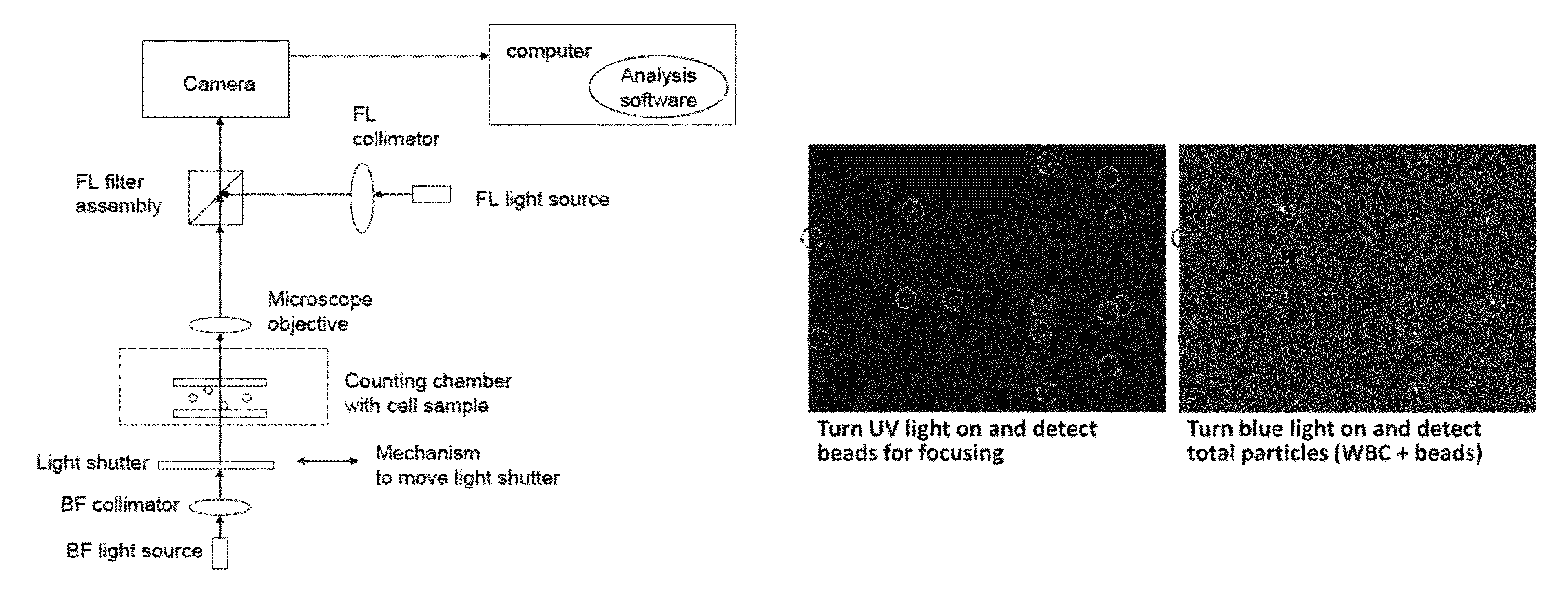
[0051]Using a large format camera, a large volume of blood was imaged, specifically for counting leukocytes for validating leukoreduced blood sample. Two images were captured (FIG. 6), one with excitation of UV and other with blue light. The blood sample was diluted 1 to 1 with Acridine Orange stain and fluorescent beads that fluoresces in both channels.

[0052]Counting only the beads in the UV excitation channel and counting all the particles in the blue excitation channel (beads+AO stained leukocytes), a subtraction result in a measurement of the leukocyte concentration in the blood sample.

example 2

Imaging of Leukocytes

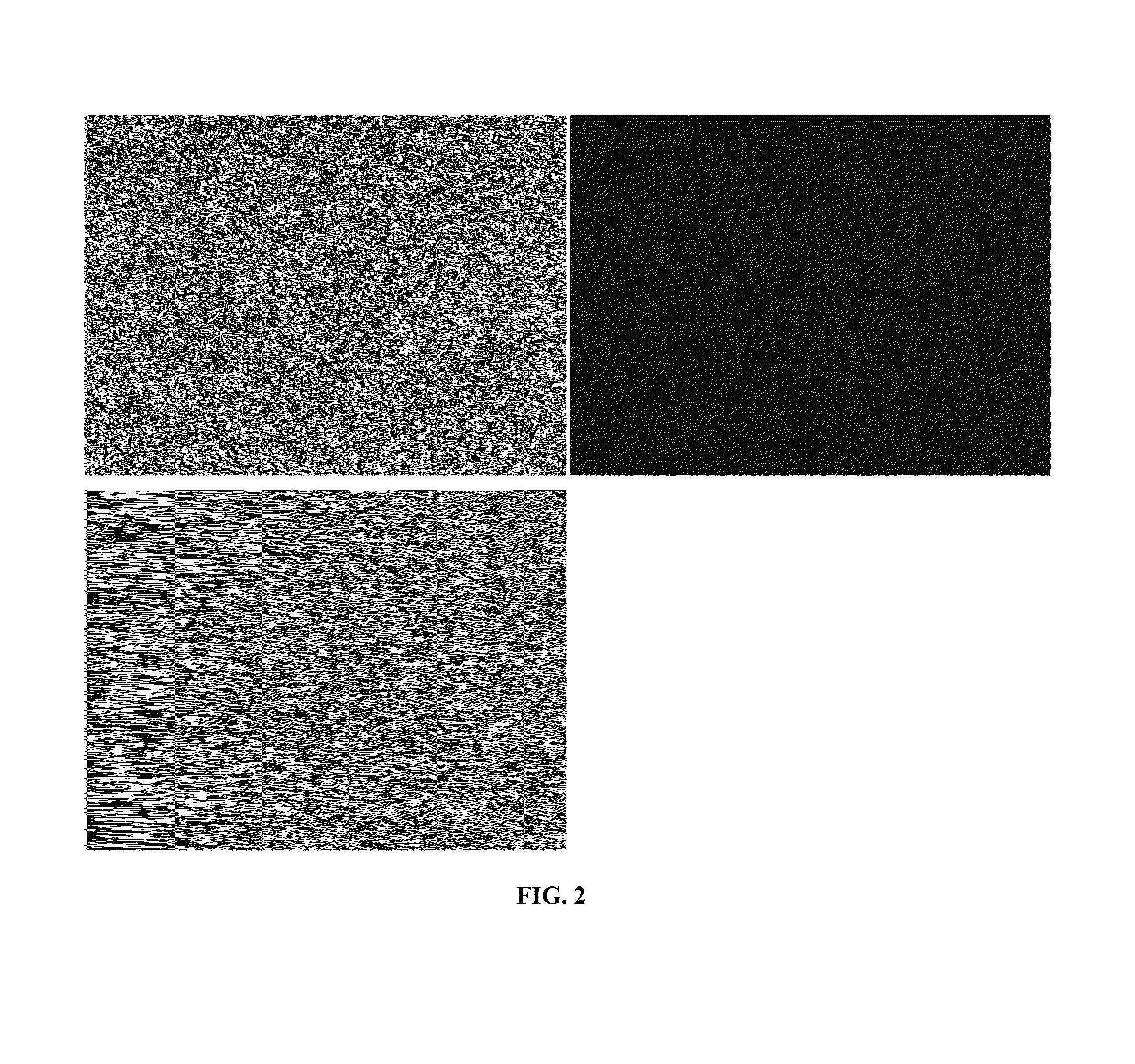
[0053]Using PI stained jurkat, with optical modules QMAX blue with 510 nm long pass filter, and 535 nm-401 nm with 510 nm long pass filter, two images were taken with UV and blue excitation (FIG. 7 and FIG. 8).

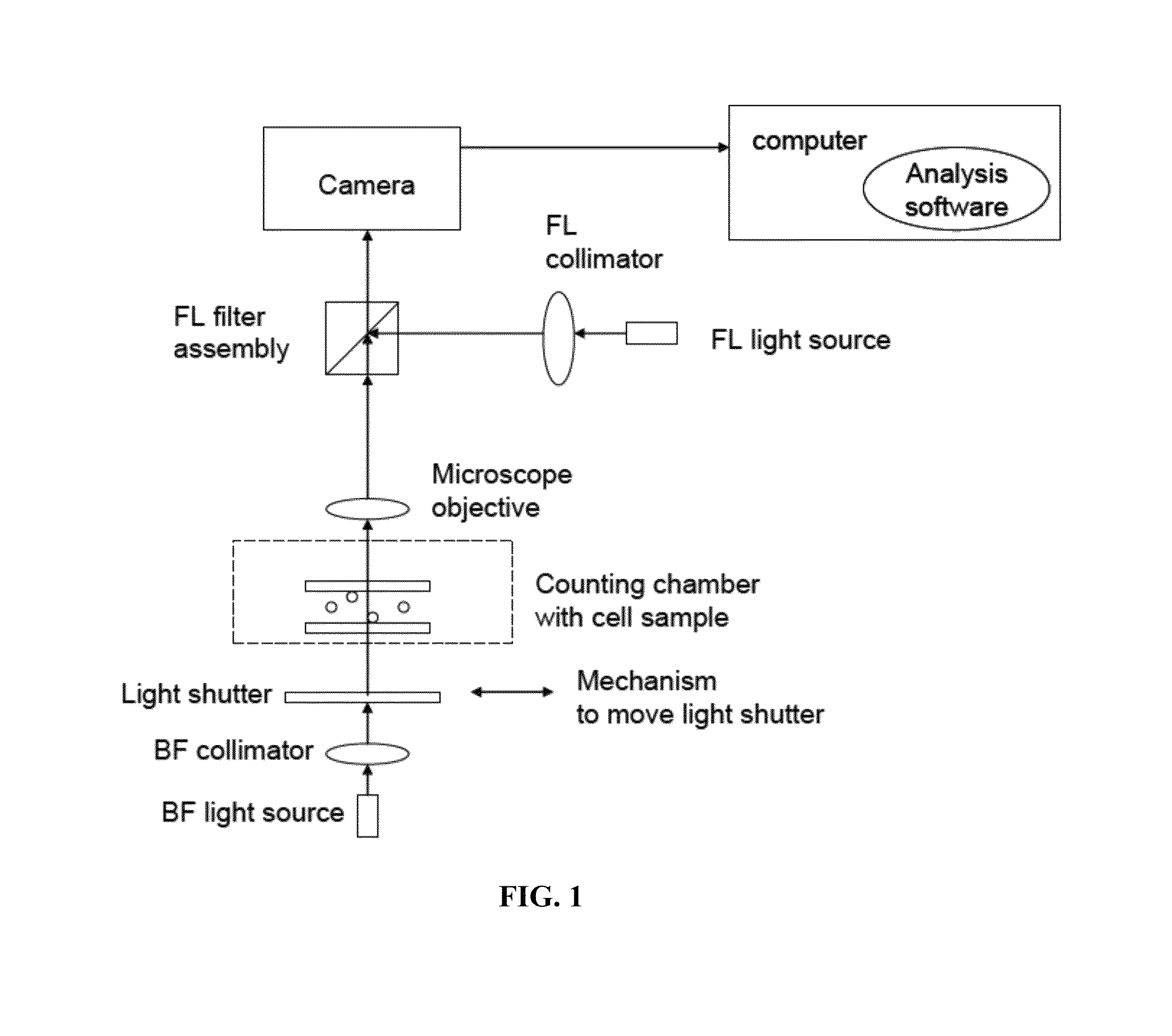
[0054]No fluorescence of beads was observed in the channel that detects PI stained Jurkats. Method would involve focusing under UV excitation, with beads, then switching to blue excitation and counting all the Cells stained with PI. This is a much simpler method than the previous described method, where a subtraction is not needed. The method is using one channel with beads to focus, and then switch to the other channel to count all the cells.


PUM

PropertyMeasurementUnit
excitation wavelengthaaaaaaaaaa
detection wavelengthaaaaaaaaaa

Abstract

The invention generally relates to analytical and monitoring systems useful for analyzing and measuring cells and biological samples. More particularly, the invention provides systems and methods for internal calibration and focus reference for cytometry imaging.

Description

PRIORITY CLAIMS AND RELATED APPLICATIONS[0001]This application claims the benefit of priority from PCT / US11 / 57177, filed Oct. 20, 2011, which claims the benefit of priority from U.S. Provisional Application Ser. No. 61 / 405,479, filed Oct. 21, 2010, the entire content of each of which is incorporated herein by reference for all purposes.TECHNICAL FIELDS OF THE INVENTION[0002]The invention generally relates to analytical and monitoring systems useful for analyzing and measuring cells and biological samples. More particularly, the invention relates to systems and methods for internal calibration and focus reference for imaging cytometry.BACKGROUND OF THE INVENTION[0003]An important aspect in the fields of medical diagnostics and biomedical research involves detection, identification, quantification, and characterization of various cells and biomolecules of interest through testing of biological samples such as blood, spinal fluid, cell culture and urine. Healthcare providers and biomed...

Claims


Application Information

Patent Timeline
Patent Type & Authority Patents(United States)
IPC IPC(8): G06K9/00G06F19/26G01N21/64G16B45/00
CPCG06F19/26G01N21/6456G01N21/6428G01N21/6486G16B45/00G01N33/5094
Inventor CHAN, LEO L.QIU, JEANLI, PETERFLANAGAN, KEVINSMITH, TIMOTHY
Owner NEXCELOM BIOSCIENCE LLC
Learn More