Eureka AIR delivers breakthrough ideas for toughest innovation challenges, trusted by R&D personnel around the world.

Electroencephalogram electrode unit for small animals and measurement system using the same

a technology of electroencephalogram and electrode unit, which is applied in the field of electroencephalogram electrode unit for small animals and a measurement system using the same, can solve the problems of difficult to fasten small electrodes to limited brain space, difficult to maintain low impedance generated between the scalp and the electrode, and the inability to use the large electrode disclosed in patent references 1 and 2 , to achieve the effect of easy increase of the number of electrodes for electroencephalogram measurement and the reduction of the metallic par

Inactive Publication Date: 2015-07-14
TOHOKU UNIV
View PDF17 Cites 8 Cited by
  • Summary
  • Abstract
  • Description
  • Claims
  • Application Information

AI Technical Summary

Benefits of technology

[0026]According to the electroencephalogram electrode unit for small animals of the present invention, the area of the metallic part of the electrode can be minimized, and thus the number of electrodes for electroencephalogram measurement can be increased easily.
[0027]By using the electroencephalogram electrode unit for small animals of the present invention, not only electroencephalogram measurement of small animals but also MRI measurement, f-MRI measurement, electrocardiographic measurement (ECG measurement), near infrared spectroscopy (NIRS), and intracranial measurement can be performed simultaneously.

Problems solved by technology

Meanwhile, with small animals such as rats, which have smaller brain volume than humans, large electrodes disclosed in Patent References 1 and 2 cannot be used.
With EEG for small animals, it is difficult to fasten small electrodes to limited brain space, and it is also difficult to maintain low impedance generated between the scalp and the electrodes.
Consequently, few reports have been made on simultaneous EEG and f-MRI measurement techniques for small animals due to such technical difficulties.
Furthermore, since EEG measurement in such EEG-f-MRI measurement uses a single electrode (See Non-patent Reference 1), electroencephalogram can be detected only partially and it is therefore not effective when the target of study is the entire brain.

Method used


Image

  • Electroencephalogram electrode unit for small animals and measurement system using the same
  • Electroencephalogram electrode unit for small animals and measurement system using the same
  • Electroencephalogram electrode unit for small animals and measurement system using the same

Examples


first embodiment

(First Embodiment)

[0048]As a first embodiment, an electroencephalogram electrode unit for small animals will be described.

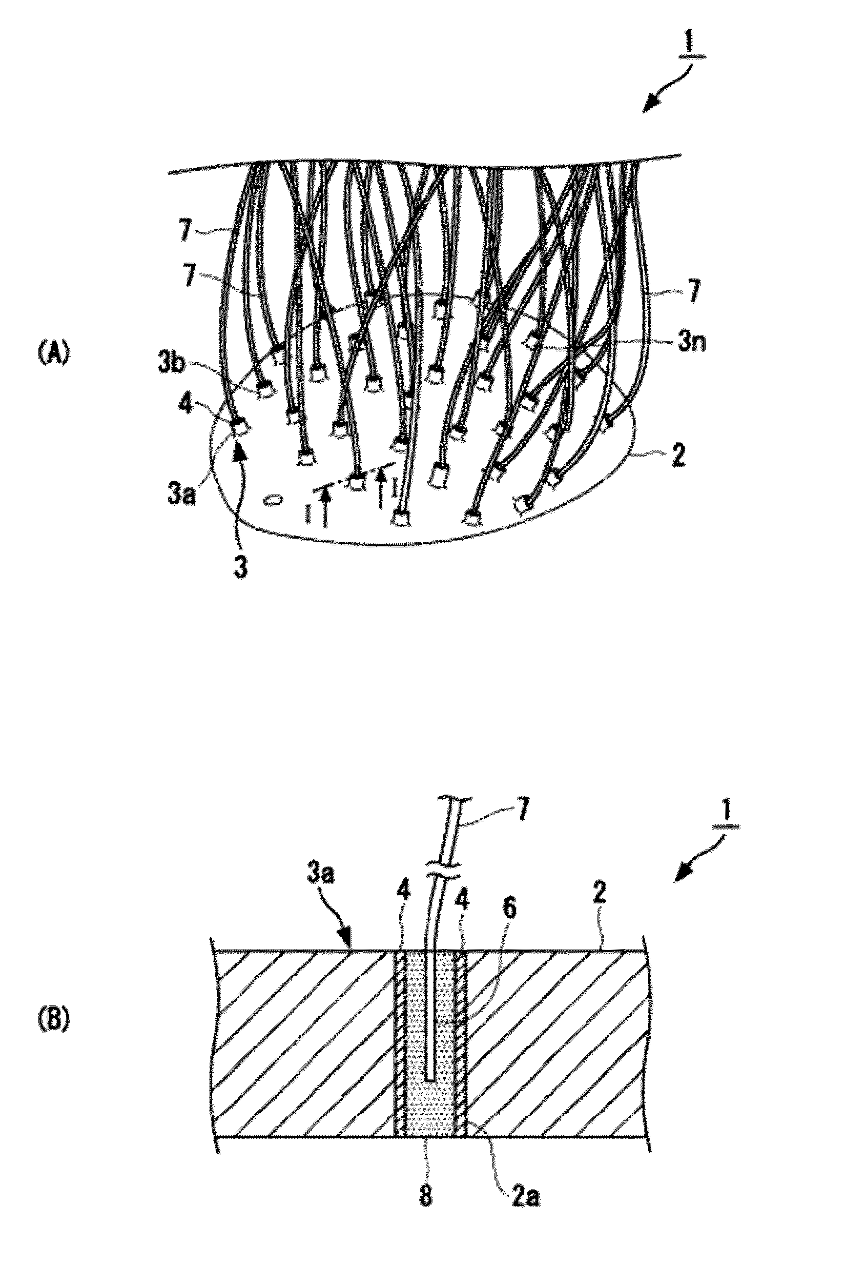
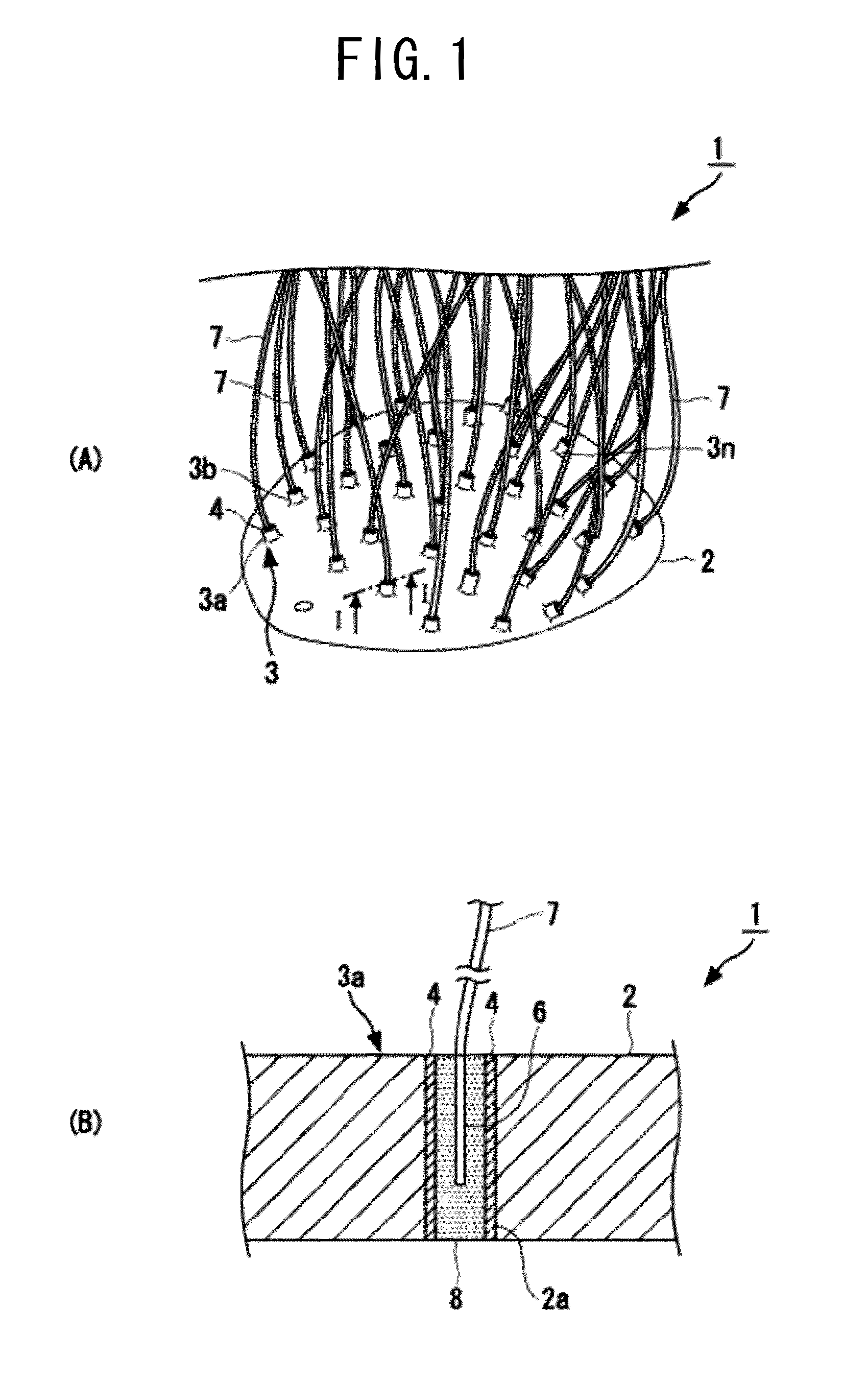
[0049]FIG. 1 shows the structure of the electroencephalogram electrode unit for small animals of a first embodiment of the present invention, wherein (A) is an oblique perspective view and (B) is a cross-sectional view along the line I-I in (A).

[0050]As shown in FIG. 1, the electroencephalogram electrode unit 1 for small animals of the present invention includes: a base 2 that covers the scalp of a small animal and has a plurality of through holes 2a; and a plurality of electrodes 3 (3a to 3n). Each of the plurality of electrodes is inserted into each of the plurality of through holes 2a. Each of the plurality of electrodes 3 is equipped with an insulating tube 4, an electrode section 6 installed within the tube 4, an extraction conducting wire 7 that is connected to the electrode section 6 and extracts electroencephalogram signals to the exterior, and a paste 8 ...


PUM


Abstract

The electroencephalogram electrode unit for small animals includes a base that covers the scalp or brain surface of a small animal and has a plurality of through holes and a plurality of electrodes. Each of the plurality of electrodes is inserted into each of the plurality of through holes, and each of the plurality of electrodes is equipped with an insulating tube, an electrode section disposed within the tube, an extraction conducting wire that is connected to the electrode section and extracts the EEG signal to outside, and a paste that is filled within the tube. The tube is installed in the through hole in a manner of standing upright from the scalp or brain surface, and the electrode section is formed in the form of a wire and is disposed, in a manner of standing upright from the scalp or brain surface, within the paste filled within the tube.

Description

TECHNICAL FIELD[0001]The present invention relates to an electroencephalogram electrode unit for small animals and a measurement system using the same. More specifically, the present invention relates to an electroencephalogram electrode unit for small animals and a measurement system using the same capable of performing electroencephalogram measurement, f-MRI and other measurements simultaneously.BACKGROUND ART[0002]Magnetic resonance imaging (MRI) is a method of imaging information within a living body using nuclear magnetic resonance phenomenon. Furthermore, the f-MRI has recently been developed. The f-MRI stands for functional magnetic resonance imaging, and is one of the methods of imaging brain functions using MRI. The changes in the neuronal activities in local areas of the brain result in the changes in the nuclear magnetic resonance imaging signals, which is the principle of the f-MRI.[0003]Furthermore, the electroencephalogram (EEG) for living bodies has conventionally bee...

Claims


Application Information

Patent Timeline
Patent Type & Authority Patents(United States)
IPC IPC(8): A61B5/0478A61B5/0408A61B5/055A61B5/296
CPCA61B5/0478A61B5/0408A61B5/04085A61B5/055A61B5/25A61B5/291A61B5/282
Inventor JORGE, RIERASUMIYOSHI, AKIRAKAWASHIMA, RYUTA
Owner TOHOKU UNIV
Why Eureka
Eureka Blog
Learn More