
Needle detection in medical image data

a technology of image data and needles, applied in the field of needle detection in medical image data, can solve the problems of inability to capture needles, difficulty in determining the tip of the needle, and difficulty in using the echo-transducer device while injecting needles, so as to reduce artifacts, simplify implementation, and achieve the effect of determining the projected needle line more accurately

Active Publication Date: 2015-09-08
KONINKLJIJKE PHILIPS NV
View PDF13 Cites 2 Cited by
  • Summary
  • Abstract
  • Description
  • Claims
  • Application Information

AI Technical Summary

Benefits of technology

[0015]Using a high-frequency linear-array transducer with frequencies of 8-17 MHz, nerves can be visualized and surrounded by local anesthetics, using a needle. This procedure results in a higher success-rate, less local anesthetics and is considered to be safer, as it reduced the risk for intravascular injection.
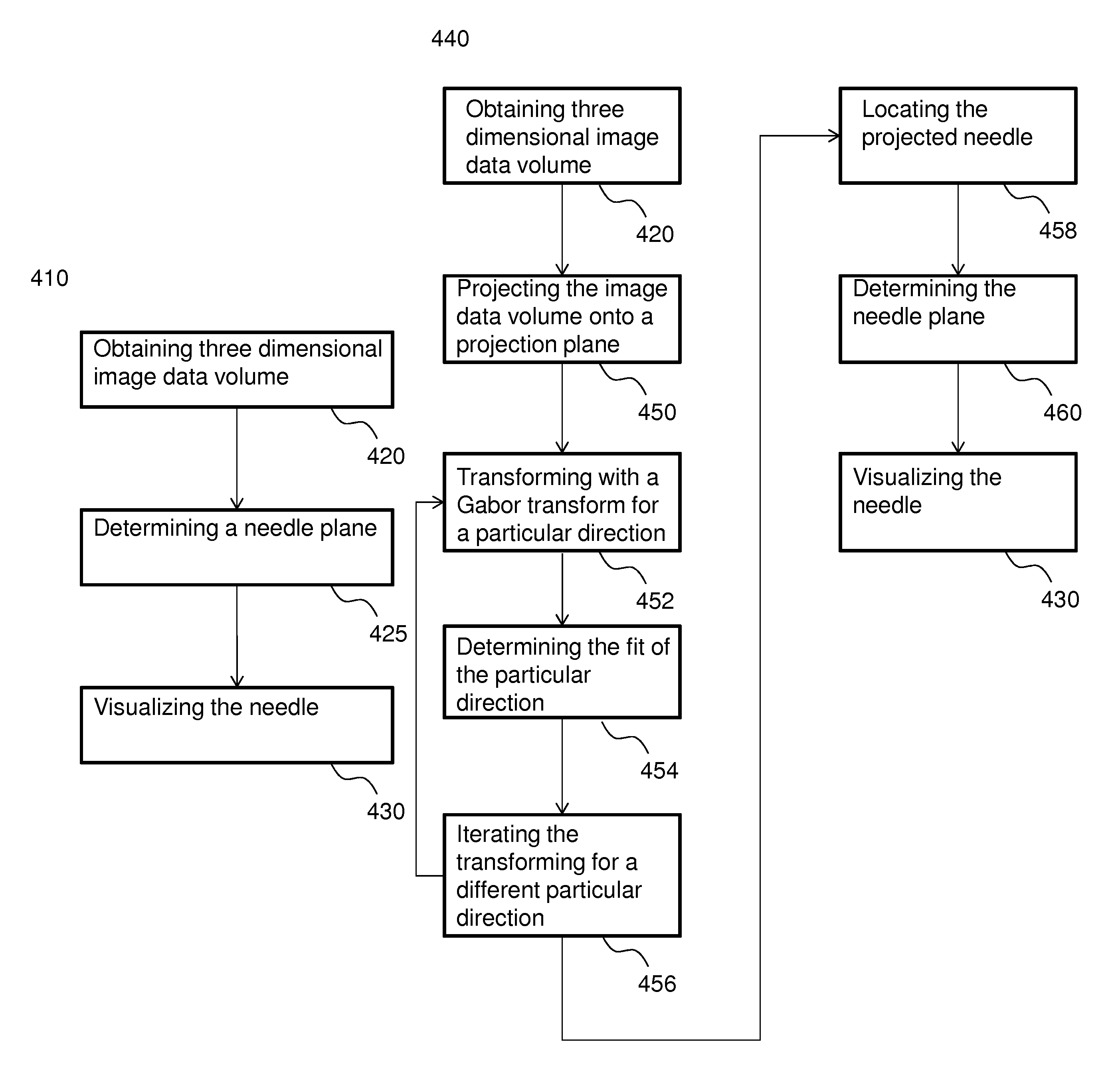
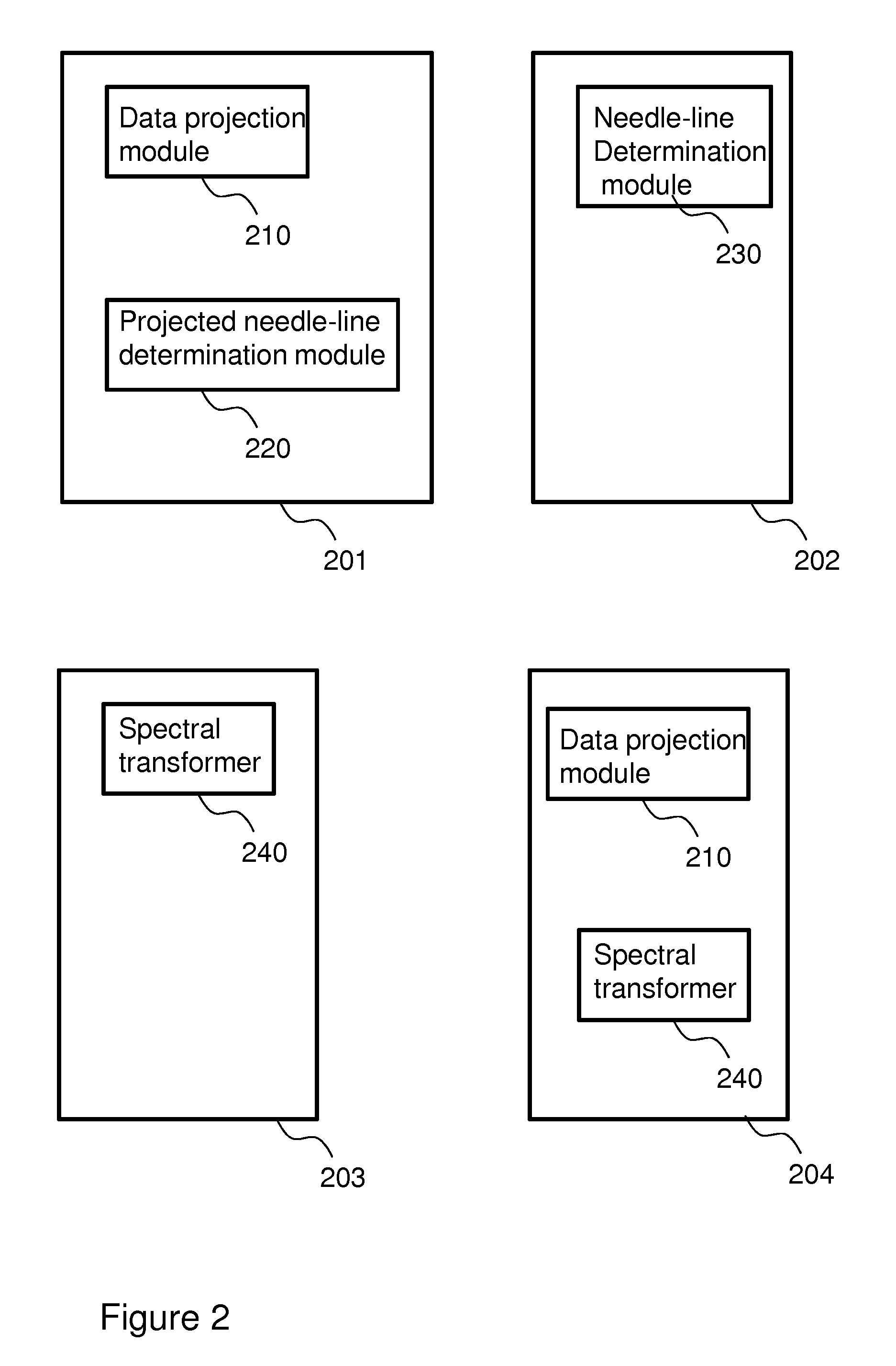
[0066]A system for processing an image data volume acquired with a medical imaging acquisition device from a body comprising a needle is provided. It has been realized it is advantageous to display a plane intersecting the image data volume showing the needle to a user of the system. This allows better positioning of the needle. The system comprises a needle-plane determination module for determining a plane being parallel to and intersecting a representation in the image data volume of the needle and being parallel to a viewing direction. The needle plane determination module may make use of pixel processing and / or spectral transformation, in particular the Gabor transform.

Problems solved by technology

It turns out however that using the echo-transducer device while injecting the needle is not so easy.
The thin needle and the thin ultrasound-field may result in failure to capture the needle.
Moreover, a physician is never sure that the tip of the needle is being visualized.
It is a problem of the prior art that echo-guided loco-regional anesthesia requires high skill from the physician in operating diagnostic sonographic scanners.
The inventors have realized that part of the problem is caused by lack of a precise alignment between the thin needle and the thin ultrasound-field.
This is very hard for the user, e.g., the physician to notice.

Method used


Image

  • Needle detection in medical image data
  • Needle detection in medical image data
  • Needle detection in medical image data

Examples


Embodiment Construction

[0124]While this invention is susceptible of embodiment in many different forms, there is shown in the drawings and will herein be described in detail one or more specific embodiments, with the understanding that the present disclosure is to be considered as exemplary of the principles of the invention and not intended to limit the invention to the specific embodiments shown and described.

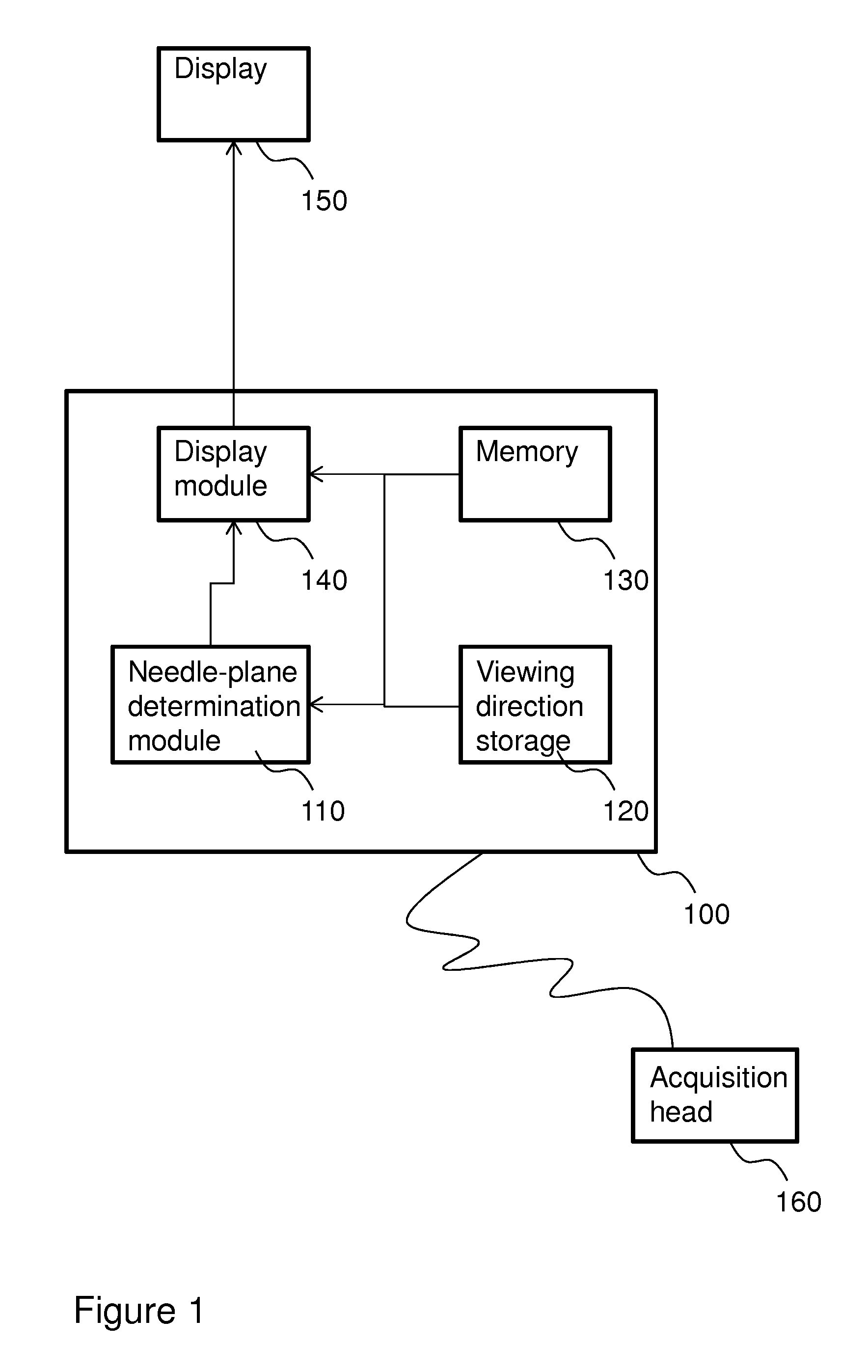
[0125]FIG. 1 shows a medical image acquisition apparatus 100. Some of the possible data dependencies between the different elements of medical image acquisition apparatus 100 are indicated in the figure with arrows.

[0126]Medical image acquisition apparatus 100 is connected, or connectable, to an image data acquisition head 160. Medical image acquisition apparatus 100 and image data acquisition head 160 together allow for the acquisition of medical image data from a body, such as a human or animal body. For example, medical image acquisition apparatus 100 may be an ultrasound apparatus and image dat...


PUM


Abstract

A system for processing an image data volume acquired with a medical imaging acquisition device 100 from a body comprising a needle is provided. It has been realized it is advantageous to display 150 a plane intersecting the image data volume showing the needle to a user of the system. This allows better positioning of the needle. The system comprises a needle-plane determination 110 module for determining a plane being parallel to and intersecting a representation in the image data volume of the needle and being parallel to a viewing direction. The needle plane determination module may make use of pixel processing and / or spectral transformation, in particular the Gabor transform.

Description

[0001]This application is the U.S. national phase of International Application No. PCT / EP2011 / 052826 filed 25 Feb. 2011 which designated the U.S. and claims priority to EP 10155335.2 filed 3 Mar. 2010, the entire contents of each of which are hereby incorporated by reference.FIELD OF THE INVENTION[0002]The invention relates to a system for processing an image data volume acquired with a medical imaging acquisition device from a body comprising a needle, the system comprising a needle-plane determination module for determining a plane being parallel to and intersecting a representation in the image data volume of the needle.[0003]The invention also relates to a medical image acquisition apparatus.[0004]The invention also relates to a method for processing an image data volume and to a corresponding computer program product and a corresponding system.BACKGROUND OF THE INVENTION[0005]For some healthcare treatments, patients are injected with a needle to transfer medication or to extrac...

Claims


Application Information

Patent Timeline
Patent Type & Authority Patents(United States)
IPC IPC(8): G06K9/00A61B8/08A61B17/34A61B19/00
CPCA61B8/0841A61B8/0833A61B8/483A61B17/3403A61B19/5244A61B2017/3413A61B2019/5265A61B2019/5276A61B34/20A61B2034/2065A61B2090/378A61B8/466A61B8/5207A61B2034/2063
Inventor KORSTEN, HENDRIKUS HUBERTUS MARIADE WITH, PETER HENDRIK NELISBERGMANS, JOHANNES WILHELMUS MARIA
Owner KONINKLJIJKE PHILIPS NV
Learn More