
Method and tomography apparatus for reconstruction of a 3D volume

a tomography and volume technology, applied in the field of 3d volume reconstruction, can solve the problems of unpredictable variations of scan geometry, non-perfect approximately circular scan, and other problems, and achieve the effect of reducing or even completely avoiding the amount of work involved

Active Publication Date: 2015-09-15
DURR DENTAL GMBH & CO KG +1
View PDF12 Cites 7 Cited by
  • Summary
  • Abstract
  • Description
  • Claims
  • Application Information

AI Technical Summary

Benefits of technology

[0011]Furthermore, it is an object of the present invention to provide a method to operate such an apparatus wherein a higher efficiency is achieved.
[0059]In this way, the time consuming optimizations can take place without interference with normal operation of the tomography apparatus. If there is only a standalone PC for data processing, control of the apparatus and presentation of the results, no extra computing power is necessary, because the demanding computations are shifted to time slots without any user interaction. It is also possible to perform more complete optimizations in such overnight time slots, which take into account more parameters or more thoroughly search the parameter space for the absolute minimum of the re-projection error, e.g. for the fine tuning of calibration parameters.

Problems solved by technology

However, because of mechanical inaccuracies of the tomography apparatus, the assumed scan geometry used during reconstruction may deviate from the actual scan geometry resulting e.g. in a non-perfect approximately circular scan.
Others might not be compensable.
For example, the tomography apparatus may be afflicted with geometrical instabilities during the scan resulting in unpredictable variations of the scan geometry.
As a result, the reconstructed 3D volume will show errors or artefacts which makes the interpretation by the user more difficult and might even lead to a wrong diagnosis.
However, it is not generally guaranteed that lower entropy corresponds to a better reconstruction.
Besides, this method only allows improvement with respect to a limited number of geometric parameters of the CT scan, whereas the degree of improvement is limited.

Method used


Image

  • Method and tomography apparatus for reconstruction of a 3D volume
  • Method and tomography apparatus for reconstruction of a 3D volume
  • Method and tomography apparatus for reconstruction of a 3D volume

Examples


Embodiment Construction

[0069]While this invention is susceptible of embodiment in many different forms, there is shown in the drawings and will herein be described in detail one or more embodiments with the understanding that the present disclosure is to be considered as an exemplification of the principles of the invention and is not intended to limit the invention to the embodiments illustrated.

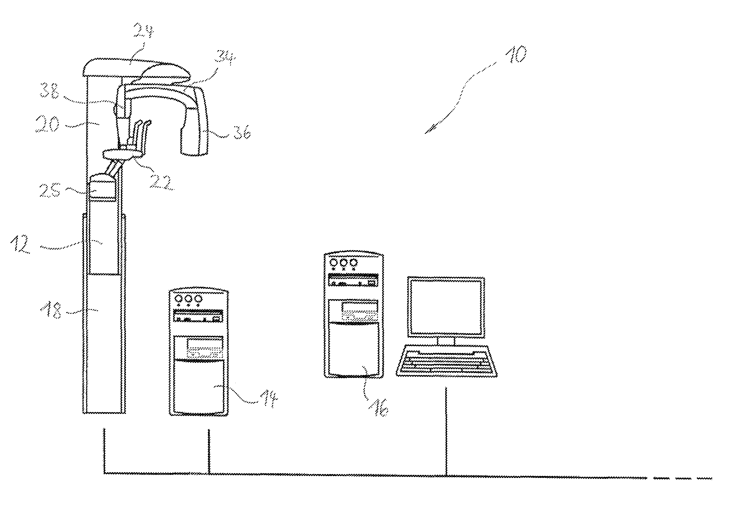
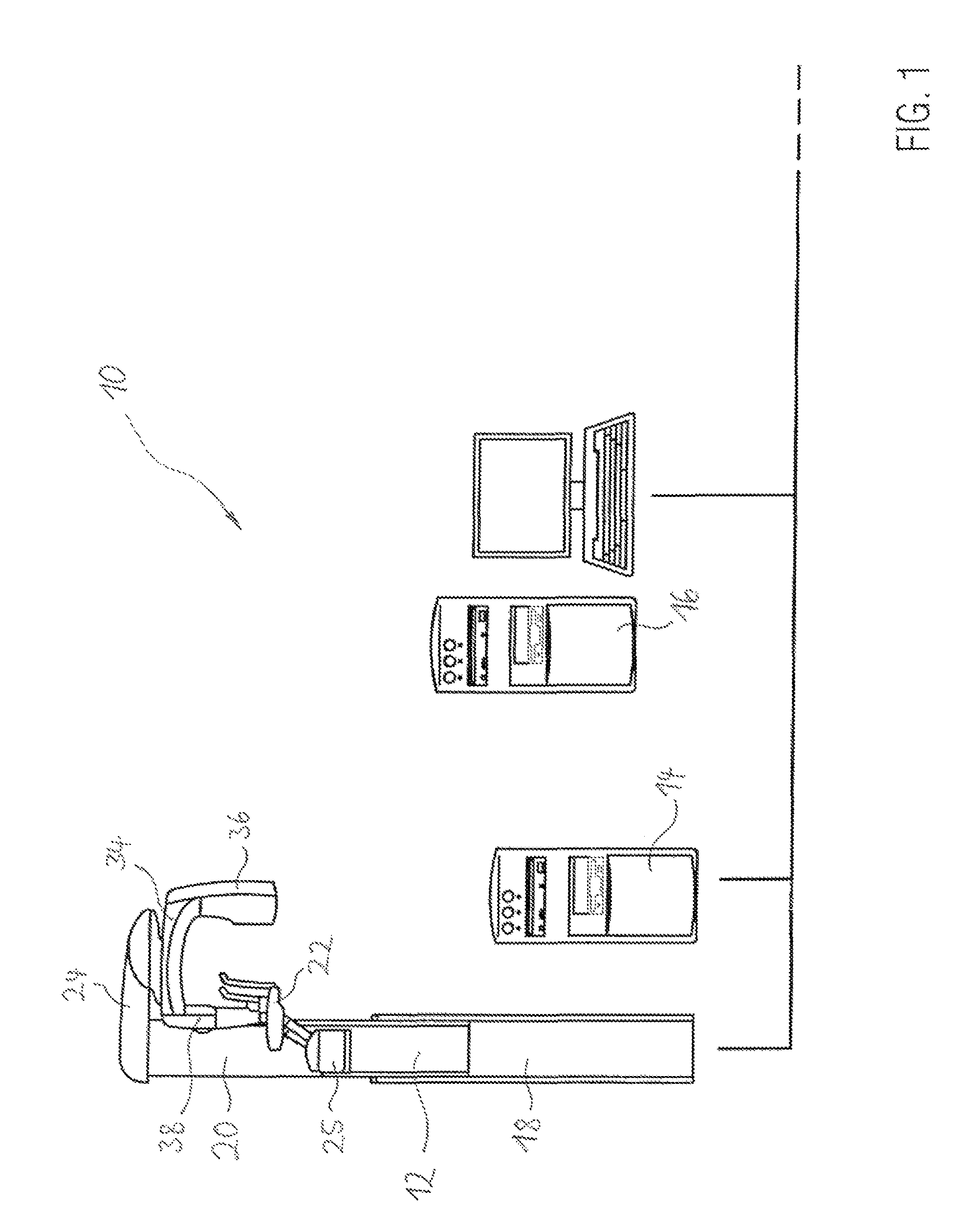
[0070]FIG. 1 shows a DVT apparatus 10 used for dental medical examinations of the jaw section of a patient comprising an acquisition unit 12, a data processing unit 14 and a workstation 16.

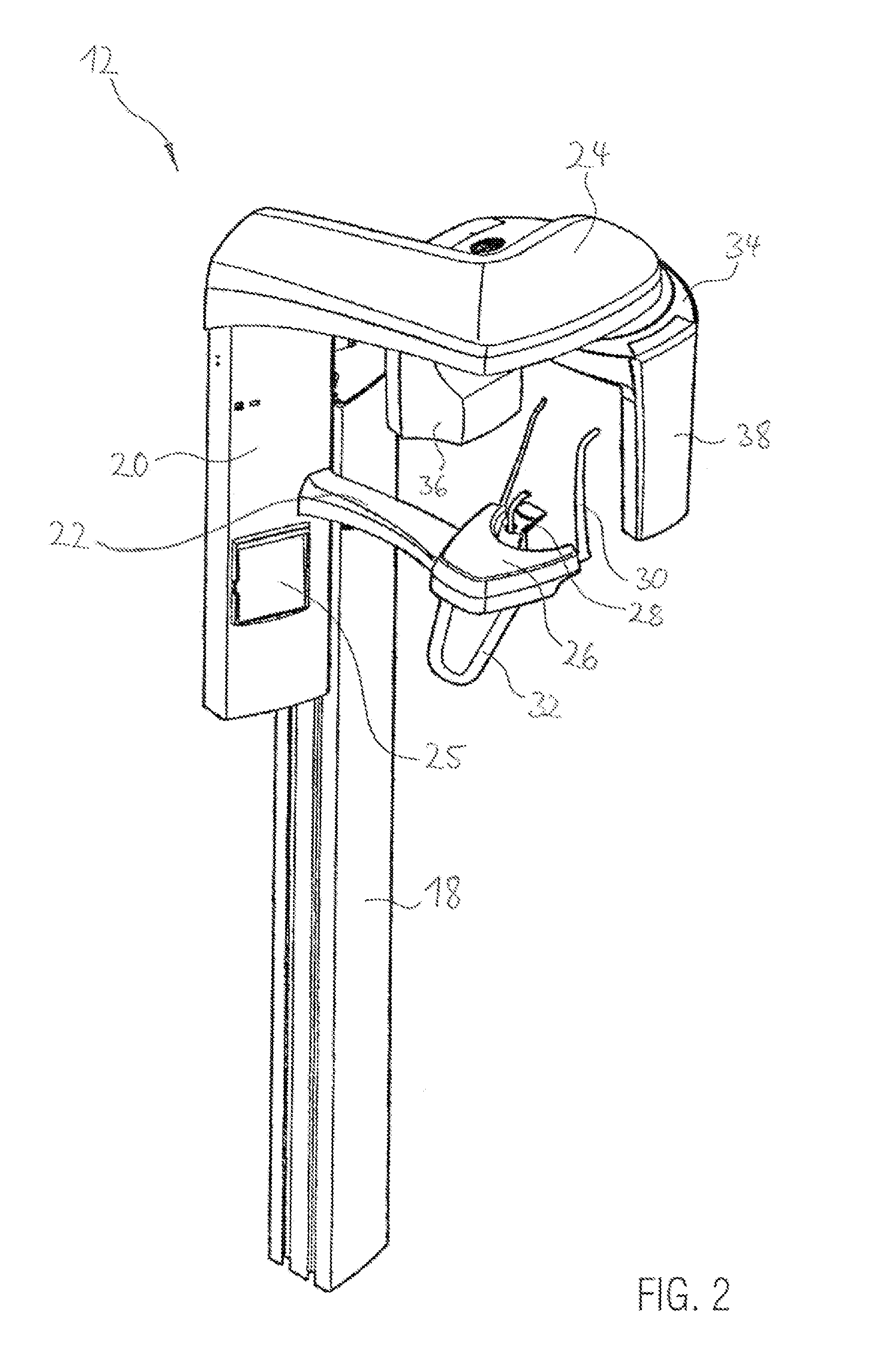
[0071]The acquisition unit 12 comprises a fixed support column 18 along which a support carrier 20 is movable in a vertical direction in order to adjust the acquisition unit 12 for different patient heights. The support carrier 20 comprises an object support beam 22 and a scanner support beam 24 which both extend horizontally from the support carrier 20. Furthermore, a control panel 25 is attached to the support carrier 20 allo...


PUM


Abstract

A method for reconstruction of a 3D volume from a set of projection images recorded by a tomography apparatus using penetrating radiation in the field of dental medical applications takes into account a given value of at least one parameter. Simulated projection images are generated which correspond to at least a subset of the recorded projection images by simulating a projection of the penetrating radiation through the reconstructed 3D volume taking into account said given value of the at least one parameter. A re-projection error is determined by comparing the simulated projection images (with the corresponding recorded projection images. The re-projection error is then minimized by changing the value of the at least one parameter and by iterating over the above steps.

Description

CROSS-REFERENCE TO RELATED APPLICATIONS[0001]The present application claims priority to European Patent Application No. 11005632.2 filed Jul. 10, 2011. The full disclosure of this earlier application is incorporated herein by reference.BACKGROUND OF THE INVENTION[0002]1. Field of the Invention[0003]The invention relates to a method for reconstruction of a 3D volume from a set of recorded projection images in the field of dental medical applications.[0004]2. Description of Related Art[0005]Computed tomography (CT) is a digital 3D imaging method using penetrating radiation in particular X-ray radiation to obtain internal 3D information of an object. In a first recording step of CT a volume at least partially including the object is examined by irradiating the volume with the penetrating radiation from a number of different directions and recording corresponding projection images.[0006]For this purpose, a tomography apparatus comprising a movable radiation source and a movable radiatio...

Claims


Application Information

Patent Timeline
Patent Type & Authority Patents(United States)
IPC IPC(8): A61B6/00G06T11/00
CPCG06T11/008A61B6/583G06T2211/424
Inventor WEIN, WOLFGANGLADIKOS, ALEXANDER
Owner DURR DENTAL GMBH & CO KG
Learn More