Medical device for treating a heart valve insufficiency or stenosis
a heart valve and insufficiency technology, applied in the field of medical devices for treating insufficiency or stenosis of heart valves, can solve the problems of inability to close, patient at considerable risk, narrowing (stenosis), etc., and achieve the effect of minimising the risk of tissue damage and the implantation process
- Summary
- Abstract
- Description
- Claims
- Application Information
AI Technical Summary
Benefits of technology
Problems solved by technology
Method used
Image
Examples
second embodiment
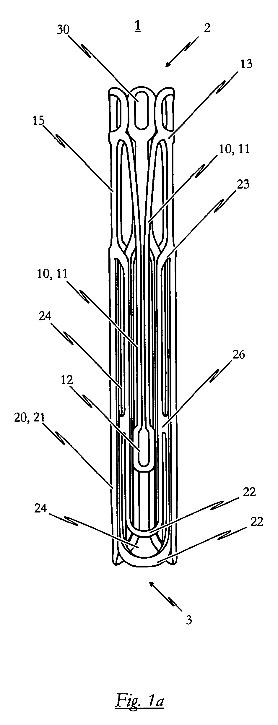
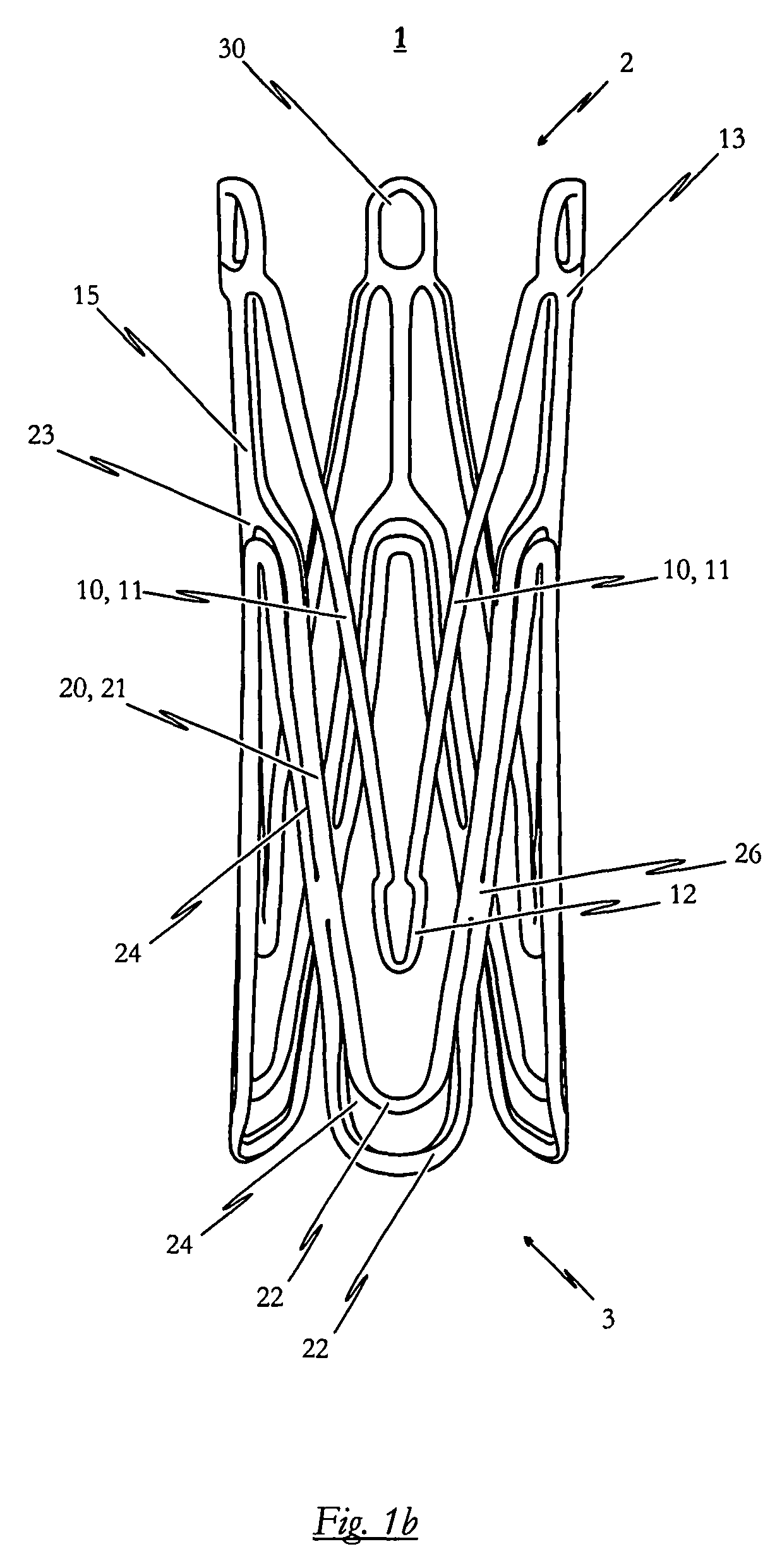
[0139]FIGS. 2a to 2c illustrate a self-expandable endoprosthesis 1 for the medical device proposed by the invention in its first, pre-definable mode (see FIG. 2a) in its second pre-definable mode (see FIG. 2c) as well as in a state in between (see FIG. 2b).
[0140]FIG. 2d illustrates a second embodiment of the medical device proposed by the invention in its expanded state with an endoprosthesis of the type illustrated in FIG. 2c and a heart valve prosthesis 40 attached to it and opened out. A flat projection of a cutting pattern which may be used for the production of the second embodiment of the self-expandable endoprosthesis is illustrated in FIG. 2e. This cutting pattern is suitable for cutting the endoprosthesis illustrated in FIG. 2a integrally from a metal tube.
[0141]The endoprosthesis 1 of the second embodiment essentially corresponds to the first embodiment described above with reference to FIGS. 1a to 1e. Description of the various components corresponds to that described for...
third embodiment
[0142]a self-expandable endoprosthesis for the medical device proposed by the invention illustrated in FIGS. 3a to 3c essentially corresponds to the first preferred embodiment illustrated in FIGS. 1a to 1c. The difference, however, is that in the third preferred embodiment, the retaining eyes 30 disposed between two adjacent positioning arches 10 are provided with barbs 17, the respective tips of which point in the direction of the proximal end 3 of the endoprosthesis 1. With this modification to the design additional anchoring is provided for the system to prevent the stent 1 from being dislocated in the direction of the left ventricle.
[0143]FIG. 3d illustrates a third embodiment of the medical device proposed by the invention in its expanded state with an endoprosthesis of the type illustrated in FIG. 3c and a heart valve prosthesis 40 attached to it and opened out. This diagram essentially corresponds to that of FIG. 1d. The difference, however, is that the barb elements 17 descr...
fifth embodiment
[0148]FIG. 5e illustrates a flat projection of a cutting pattern which may be used for production of the self-expandable endoprosthesis 1 to cut the endoprosthesis 1 illustrated in FIG. 5a integrally from a metal tube.
[0149]The sixth embodiment of the self-expandable endoprosthesis and the medical device proposed by the invention illustrated in FIGS. 6a to 6e corresponds to a combination of the second embodiment illustrated in FIGS. 2a to 2e and the fifth embodiment described above with reference to FIGS. 5a to 5e. Specifically, therefore, the endoprosthesis 1 based on the second embodiment is provided with additional reinforcing portions 26 at the respective retaining arches 21 which interrupt the slots 24 extending in the longitudinal direction of the retaining arches 21.
[0150]The seventh embodiment of the endoprosthesis 1 and the medical device proposed by the invention illustrated in FIGS. 7a to 7e corresponds to a combination of the third and fifth embodiments described above. ...
PUM
 Abstract
Description
Claims
Application Information
 - R&D
- Intellectual Property
- Life Sciences
- Materials
- Tech Scout
- Unparalleled Data Quality
- Higher Quality Content
- 60% Fewer Hallucinations
Browse by: Latest US Patents, China's latest patents, Technical Efficacy Thesaurus, Application Domain, Technology Topic, Popular Technical Reports.
© 2025 PatSnap. All rights reserved.Legal|Privacy policy|Modern Slavery Act Transparency Statement|Sitemap|About US| Contact US: help@patsnap.com


