

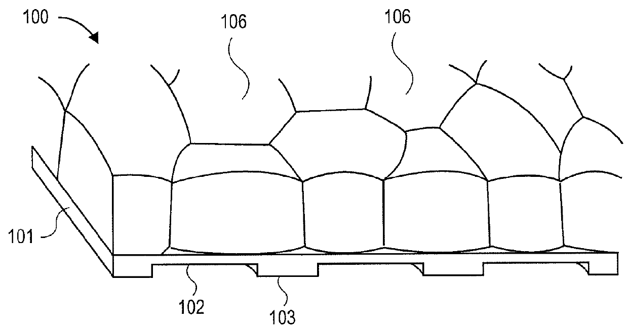
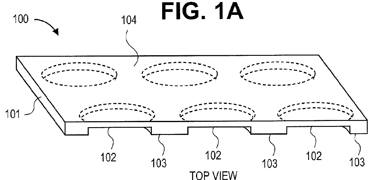
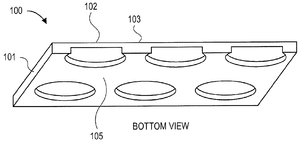
3-Dimensional parylene scaffold cage
a parylene scaffold and cage technology, applied in the field of 3-dimensional parylene scaffold cages, to achieve the effect of hindering or preventing cell migration
- Summary
- Abstract
- Description
- Claims
- Application Information
AI Technical Summary
Benefits of technology
Problems solved by technology
Method used
Image

Examples
example 1
[0108]Illustrates the Permeability of the Implantable Cage-Like Apparatus is Comparable to a Bruch's Membrane
[0109]To measure the permeability of the bottom substrate, a blind-well diffusion experiment was performed. A mesh-supported submicron parylene-C with 0.3 μm submicron membrane was clamped in-between two blind well chambers (Neuro Probe, Inc.). FITC-dextran molecules (Sigma-Aldrich Co.) with various molecular weights (MW) were initiated in the bottom chamber. The top chamber was initially filled with phosphate buffered saline (PBS).
[0110]By monitoring the fluorescence intensity in the top chamber, the diffusion coefficients of dextran molecules were calculated using a previously described method (B. Lu, Z. Liu and Y. C. Tai, “Ultrathin parylene-C semipermeable membranes for biomedical applications”, Proc. of MEMS 2011, Cancun, Mexico, Jan. 23-27, 2011, pp. 505-508). The permeability of 0.3 μm parylene-C was compared with the healthy human Bruch's membranes, in terms of the mo...
example 2
Illustrates the Mechanical Strength of the Implantable Apparatus
[0115]Example 2 illustrates membrane deflection experiments to evaluate the mechanical properties of the parylene membranes, in terms of their yielding pressures and breakdown pressures. The yielding pressure was defined as the minimum pressure that could create wrinkles and irreversible deformation. The breakdown pressure was recorded at the moment the membrane broke. Compared to the large uniform 0.3 μm parylene film, it was observed that the mesh-supported structure greatly improved the mechanical strength of the entire membrane (Table 2).
[0116]
TABLE 2Membrane typesRatio ofYieldingBreaking(overall diameter is 1.8 mm)thin partpressurepressureUniform 0.3 μm parylene100%1.50 psi 3.32 psiMesh-supported 0.3 μm 53%4.88 psi19.94 psiparylene
[0117]FIG. 16A shows the membrane mechanical property testing experimental set-up. As shown therein, the membrane pressure deflection testing set-up includes a microscope; an exposed memb...
example 3
Illustrates a Cell Migration Experiment of the Implantable Apparatus
[0119]Once sealed inside a cage, RPE cells need to develop microvilli to connect to photoreceptors. Therefore, the cage's top cover can have a fishnet-like structure, exposing the microvilli, but blocking the migration of the whole cell. In one embodiment, the gap between the top and bottom was 6 μm, determined by the height of adhering polarized cells.
[0120]To determine the size of opening, cell migration experiments were carried out with the transwell setup. Porous parylene membranes with 8 μm-diameter and 4 μm-diameter holes were fabricated using the method described in (B. Lu, T. Xu, S. Zheng, A. Goldkorn and Y. C. Tai, “Parylene membrane slot filter for the capture, analysis and culture of viable circulating tumor cells”, Proc. of MEMS 2010, Hong Kong, China, Jan. 24-28, 2010, pp. 935-938). Platelet-derived growth factor (PDGF) was added to the bottom well, which was a stimulus of cell migration. Holes with 8 μ...
PUM
| Property | Measurement | Unit |
|---|---|---|
| molecular weight | aaaaa | aaaaa |
| molecular weight | aaaaa | aaaaa |
| thick | aaaaa | aaaaa |
Abstract
Description
Claims
Application Information