Segmented scaffolds and delivery thereof for peripheral applications

a technology of polymer scaffolds and peripheral vessels, applied in the field of stent scaffolds, can solve the problems of affecting the delivery of polymer scaffolds in peripheral vessels, affecting the treatment of blood vessels, and affecting the effect of restnosis

Inactive Publication Date: 2016-02-09
ABBOTT CARDIOVASCULAR
View PDF102 Cites 3 Cited by
  • Summary
  • Abstract
  • Description
  • Claims
  • Application Information

AI Technical Summary

Benefits of technology

[0017]Embodiments of the present invention include a method for delivering a segmented scaffold comprising: providing a delivery balloon comprising a plurality of disconnected scaffold segments arranged end to end such that adjacent segments are spaced apart by a gap, wherein the segments are crimped to the balloon; and inflating the balloon to radially expand the segments, wherein the balloon shortens as it is inflated, wherein the shortening of the balloon reduces the increase in the gap between adjacent segments caused by shortening of the segments as they expand
[0026]Embodiments of the present invention include a method of delivering a segmented scaffold comprising: providing a delivery balloon, wherein two disconnected scaffold segments arranged end to end and spaced apart by a gap are disposed over the balloon, wherein the segments are crimped to the balloon; inflating and expanding the balloon to expand the segments, wherein expansion of the segments increases the width of the gap between the segments; and applying an axially directed force to the balloon to shorten the balloon to reduce the width of the gap.
[0027]Embodiments of the present invention include a system for deployment of a segmented scaffold comprising: an outer tubular member; an inner elongate member disposed within the outer tubular member; and a delivery balloon disposed over at least the inner elongate member, wherein a proximal end of the balloon is attached to the outer tubular member and a distal end of the balloon is attached to a distal end of the inner elongate member, wherein the inner elongate member is axially slideable within the outer tubular member, wherein when the balloon is at least partially inflated, sliding the inner elongate member proximally causes the balloon to shorten.
[0030]Embodiments of the present invention include a method for delivering a segmented scaffold comprising: positioning a delivery balloon comprising a plurality of disconnected scaffold segments arranged end to end and spaced apart by gaps at a treatment site within a blood vessel of a patient, wherein the segments are crimped to the balloon, wherein a surface of the balloon between the gaps comprises a coating including a therapeutic agent; and inflating the balloon to radially expand and implant the segments at the implant site, wherein the therapeutic agents reduces restenosis at the implant site at or proximal to the gaps.

Problems solved by technology

Because these stents are close to the surface of the body, they are particularly vulnerable to crushing or pinching loads, which can partially or completely collapse the stent and thereby block fluid flow in the vessel.
However, in many treatment applications, the presence of a stent in a body is necessary for a limited period of time until its intended function of, for example, maintaining vascular patency and / or drug delivery is accomplished.
However, there are numerous challenges to overcome when developing a polymer scaffold, particularly in peripheral blood vessels, or blood vessels outside the coronary arteries in which a stent is subjected to both radial forces and nonpulsatile forces.
Additionally, there are challenges in delivery of polymer scaffolds in peripheral vessels.

Method used


Image

  • Segmented scaffolds and delivery thereof for peripheral applications
  • Segmented scaffolds and delivery thereof for peripheral applications
  • Segmented scaffolds and delivery thereof for peripheral applications

Examples


example 1

Pillowed Balloon to Set Segment Spacing

[0236]FIG. 21A depicts a photograph of a delivery balloon in a deflated state with pre-pillowed bands or sections to set scaffold segment spacing.

[0237]FIG. 21B depicts a close-up view of a pre-pillowed section. With the pre-pillowing of the balloon, the segments not only maintain a consistent spacing during crimping, but also when deployed.

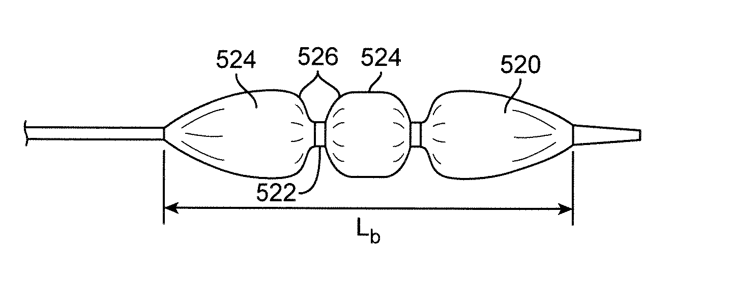
[0238]FIG. 22 illustrates the inflation process of a segmented scaffold that shows that even segment spacing is maintained throughout expansion of the segments. The top picture shows a segmented scaffold crimped over a deflated balloon. The middle picture depicts the expansion when the balloon is partially inflated with the balloon having a dog bone shape. The ends of the balloon are inflated which partially expands the segments at the ends. The center of the balloon is partially or not inflated. The bottom picture shows the completely inflated balloon and the segments completely expanded. The balloon pre-pi...

example 2

Animal Studies of inflation of balloon with pre-pillowed sections

[0239]An animal study of the pre-pillowed balloon inflation with a segmented scaffold was performed using a porcine model. A balloon with pre-pillowed sections with a segmented scaffold first inflated at the ends creating a dog bone shape and then in the center of the balloon. The balloon shape change was observed by the contrasts shape under fluoroscopy. The final inflated balloon image showed even spacing of the implanted scaffold segments.

[0240]FIG. 23A is a fluoroscopic image depicting implanted scaffold segments. FIG. 23A depicts the segmented scaffold implanted in the Right External Iliac in a porcine model. The even spacing of the radiopaque markers is shown by the arrows. The polymer scaffold does not show up under fluoroscopy so equal distance between markers signifies equal segment spacing. FIG. 23B is a close-up view of a segment which shows the radiopaque markers.

example 3

Pre-Crimping and Final Crimping of Scaffold Segments on a Pre-Pillowed Balloon

[0241]FIG. 24A depicts scaffold segments placed on a stepped mandrel for loading into a pre-crimper.

[0242]FIG. 24B depicts pre-crimped segments of FIG. 24A upon removal from the pre-crimper.

[0243]FIG. 25A shows pre-crimped segments of FIG. 24B loaded onto a pre-pillowed balloon.

[0244]FIG. 25B shows the finished crimped segments of FIG. 25A.

[0245]FIG. 25C is a close up view of the final crimped scaffold of FIG. 25B showing pillowing between the segments.


PUM


Abstract

Segmented scaffolds composed of disconnected scaffold segments are delivered to an implant site on a delivery balloon. The segments are crimped to the balloon and separated from each other by gaps. When the balloon is inflated the gaps between the disconnected scaffold segments stays constant Pillowed or banded balloons are used to deliver the segments.

Description

BACKGROUND OF THE INVENTION[0001]1. Field of the Invention[0002]This invention relates to methods of treatment of blood vessels with metallic and polymeric medical devices, in particular, stent scaffolds.[0003]2. Description of the State of the Art[0004]This invention relates to radially expandable endoprostheses, that are adapted to be implanted in a bodily lumen. An “endoprosthesis” corresponds to an artificial device that is placed inside the body. A “lumen” refers to a cavity of a tubular organ such as a blood vessel. A stent is an example of such an endoprosthesis. Stents are generally cylindrically shaped devices that function to hold open and sometimes expand a segment of a blood vessel or other anatomical lumen such as urinary tracts and bile ducts. Stents are often used in the treatment of atherosclerotic stenosis in blood vessels. “Stenosis” refers to a narrowing or constriction of a bodily passage or orifice. In such treatments, stents reinforce body vessels and prevent r...

Claims


Application Information

Patent Timeline
Patent Type & Authority Patents(United States)
IPC IPC(8): A61F2/06A61F2/958A61F2/966A61F2/82
CPCA61F2/958A61F2/966A61F2002/826A61F2002/9583A61F2210/0076A61F2250/0019A61F2250/0039A61F2/844A61F2/91A61F2/915
Inventor PAPP, JOHN E.NGUYEN, HUNG T.ELERATH, JUSTIN K.HERABADI, DION I.MARTINS, MICHAEL T.KING, KIRSTEN S.MUELLER, SCOTT H.PELOQUIN, COETA K.PACETTI, STEPHEN D.
Owner ABBOTT CARDIOVASCULAR
Why Eureka
Try Eureka