
Volume image reconstruction using data from multiple energy spectra

a technology of multiple energy spectra and volume image, applied in the field of radiographic imaging, can solve the problems of compromising image quality, requiring an expensive detector capable of carrying out both, and conventional digital radiography detectors have limitations related to the attenuation of radiation energy, so as to reduce exposure levels

Active Publication Date: 2016-02-23
CARESTREAM DENTAL TECH TOPCO LTD
View PDF8 Cites 25 Cited by
  • Summary
  • Abstract
  • Description
  • Claims
  • Application Information

AI Technical Summary

Benefits of technology

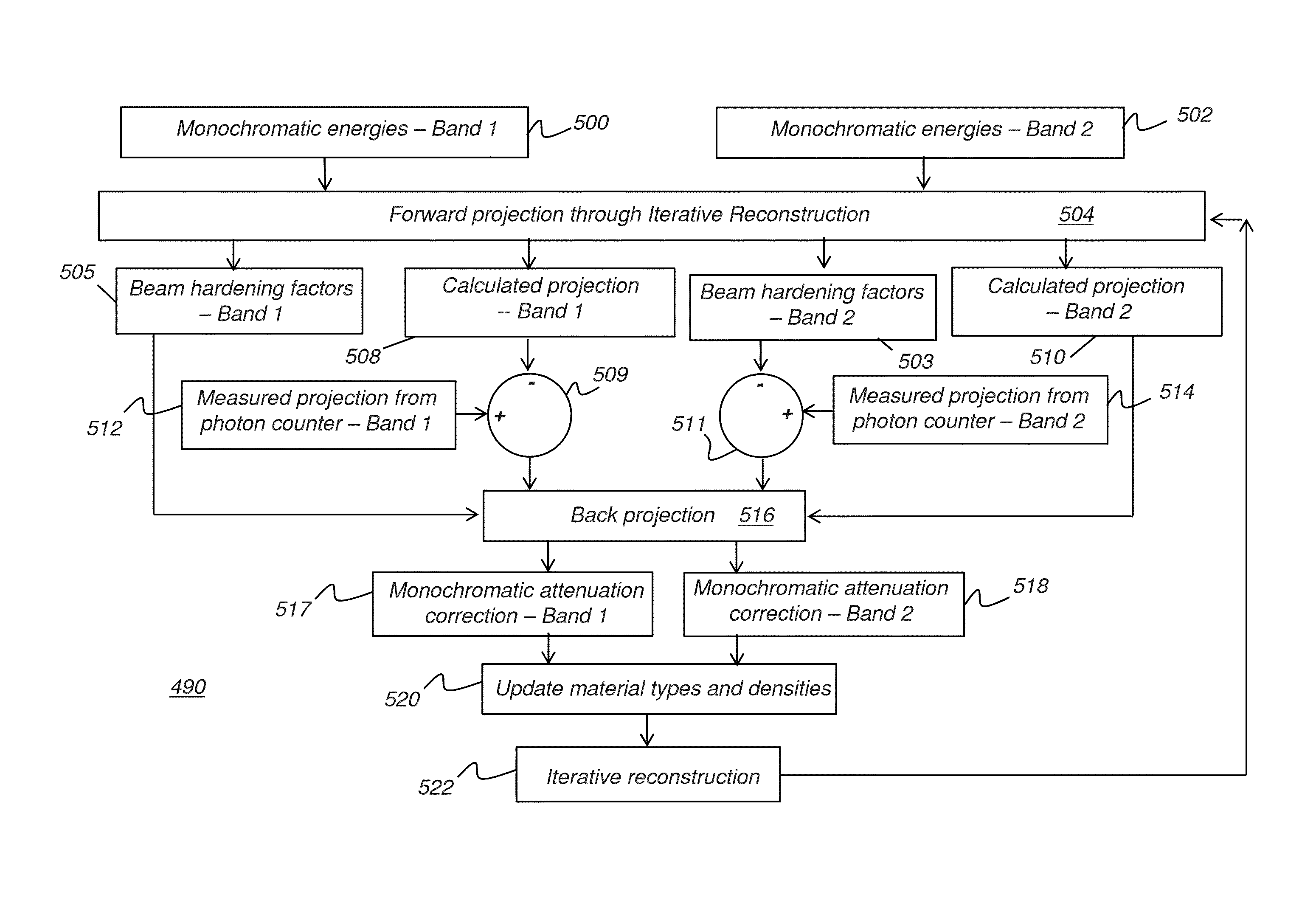
The present invention is about improving the imaging of heads and dental applications. It uses a method called photon-counting to get useful images using reduced exposure levels. The invention also includes an iterative reconstruction method to reduce beam hardening artifacts in cone beam CT reconstruction. This method helps to identify the material composition of the scanned object by transforming the X-ray attenuation coefficient from polychromatic to monochromatic values at one or more reference monochromatic energies. Overall, the invention provides better image quality for dental and ENT applications.

Problems solved by technology

However, the proposed imaging apparatus requires an expensive detector capable of carrying out both imaging functions in a satisfactory manner.
Additionally, systems of this type typically compromise image quality by using a uniform distance between the X-ray source and detector, even though different distances would be more advantageous.
Conventional digital radiography detectors have some limitations related to how attenuation of radiation energy at a single exposure is interpreted.
For example, it can be very difficult, from a single exposure, to distinguish whether an imaged object has a given thickness or a given attenuation coefficient.
This problem can be compounded for CT imaging, in which multiple images are obtained, one from each of a number of angles of revolution about the patient.
Among the problems encountered in obtaining image data for accurate 3D reconstruction is beam hardening.
This change in the spectral content of the beam causes artifacts such as cupping, in which the middle of the subject experiences different radiation levels than portions of the edge of the subject.
Beam hardening complicates the task of 3D image reconstruction in CBCT and other volume imaging modalities.

Method used


Image

  • Volume image reconstruction using data from multiple energy spectra
  • Volume image reconstruction using data from multiple energy spectra
  • Volume image reconstruction using data from multiple energy spectra

Examples


Embodiment Construction

[0044]The following is a description of exemplary embodiments of the invention, reference being made to the drawings in which the same reference numerals identify the same elements of structure in each of the several figures.

[0045]In the context of the present disclosure, the terms “pixel” and “voxel” may be used interchangeably to describe an individual digital image data element, that is, a single value representing a measured image signal intensity. Conventionally an individual digital image data element is referred to as a voxel for 3-dimensional volume images and a pixel for 2-dimensional images. Volume images, such as those from CT or CBCT apparatus, are formed by obtaining multiple 2-D images of pixels, taken at different relative angles, then combining the image data to form corresponding 3-D voxels. For the purposes of the description herein, the terms voxel and pixel can generally be considered equivalent, describing an image elemental datum that is capable of having a ran...


PUM


Abstract

A method for forming a three-dimensional reconstructed image acquires two dimensional measured radiographic projection images over a set of projection angles, wherein the measured projection image data is obtained from an energy resolving detector that distinguishes first and second energy bands. A volume reconstruction has image voxel values representative of the scanned object by back projection of the measured projection data. Volume reconstruction values are iteratively modified to generate an iterative reconstruction by repeating, for angles in the set of projection angles and for each of a plurality of pixels of the detector: generating a forward projection that includes calculating an x-ray spectral distribution at each volume voxel, calculating an error value by comparing the generated forward projection value with the corresponding measured projection image value, and adjusting one or more voxel values using the calculated error value and the x-ray spectral distribution. The generated iterative reconstruction displays.

Description

FIELD OF THE INVENTION[0001]The invention relates generally to the field of radiographic imaging and more particularly to apparatus and methods for obtaining volume images of a patient, such as images of the head.BACKGROUND OF THE INVENTION[0002]A computerized tomography (CT) imaging apparatus operates by acquiring multiple 2D images with a rotating imaging ensemble or gantry that has an x-ray source and, opposite the x-ray source, an imaging sensor rotating about a fixed axis relative to the patient. CT imaging allows the reconstruction of 3D or volume images of anatomical structures of the patient and is acknowledged to be of particular value for obtaining useful information for assisting diagnosis and treatment.[0003]There is considerable interest in the use of CT imaging in dental and ear-nose-throat (ENT) applications, as well as for other imaging of the patient's head. A number of volume imaging system designs have been proposed for this purpose. Among proposed solutions are h...

Claims


Application Information

Patent Timeline
Patent Type & Authority Patents(United States)
IPC IPC(8): G06T11/00A61B6/00A61B6/03A61B6/14A61B6/51
CPCG06T11/006A61B6/4241A61B6/482A61B6/501A61B6/5205A61B6/032A61B6/14A61B6/4085A61B6/4441A61B6/5258G06T2211/408G06T2211/424A61B6/51
Inventor INGLESE, JEAN-MARCSCHILDKRAUT, JAY S.
Owner CARESTREAM DENTAL TECH TOPCO LTD
Learn More