
Digital image analysis of inflammatory cells and mediators of inflammation

Active Publication Date: 2016-03-29
FLAGSHIP BIOSCI
View PDF32 Cites 27 Cited by
  • Summary
  • Abstract
  • Description
  • Claims
  • Application Information

AI Technical Summary

Benefits of technology

The patent describes new methods for studying inflammatory cells and their impact on cancer. These methods involve analyzing tissue sections and extracting information from them using software. The system can detect and characterize specific types of inflammatory cells and determine the overall state of the immune system in the tissue. This information can then be used to evaluate and treat patients with cancer. These methods are more advanced than current methods and can provide more accurate data.

Problems solved by technology

However, the intricate spatial relationships and the often complex distribution of inflammatory cells in tissues pose significant challenges for manual evaluation of tissue sections.
Manual evaluation of histologic tissue sections by a pathologist can be limited in several ways: (i) manual counting of inflammatory cells and determining expression levels of inflammation modulators are subjective and prone to observer bias and human error; (ii) a manual evaluation cannot practically assess a whole tissue section; (iii) quantitation of complex inflammation modulator distribution metrics (i.e. distances between cells, distances between cells and tissue features, fractal patterns, lacunarity, etc.) are not possible; and (iv) quantitative assessments of associations between one or more markers of inflammatory cell types using serial tissue sections are not possible by manual counting.

Method used


Image

  • Digital image analysis of inflammatory cells and mediators of inflammation
  • Digital image analysis of inflammatory cells and mediators of inflammation
  • Digital image analysis of inflammatory cells and mediators of inflammation

Examples


Embodiment Construction

[0028]In the following description, for purposes of explanation and not limitation, details and descriptions are set forth in order to provide a thorough understanding of the present invention. However, it will be apparent to those skilled in the art that the present invention may be practiced in other embodiments that depart from these details and descriptions without departing from the spirit and scope of the invention.

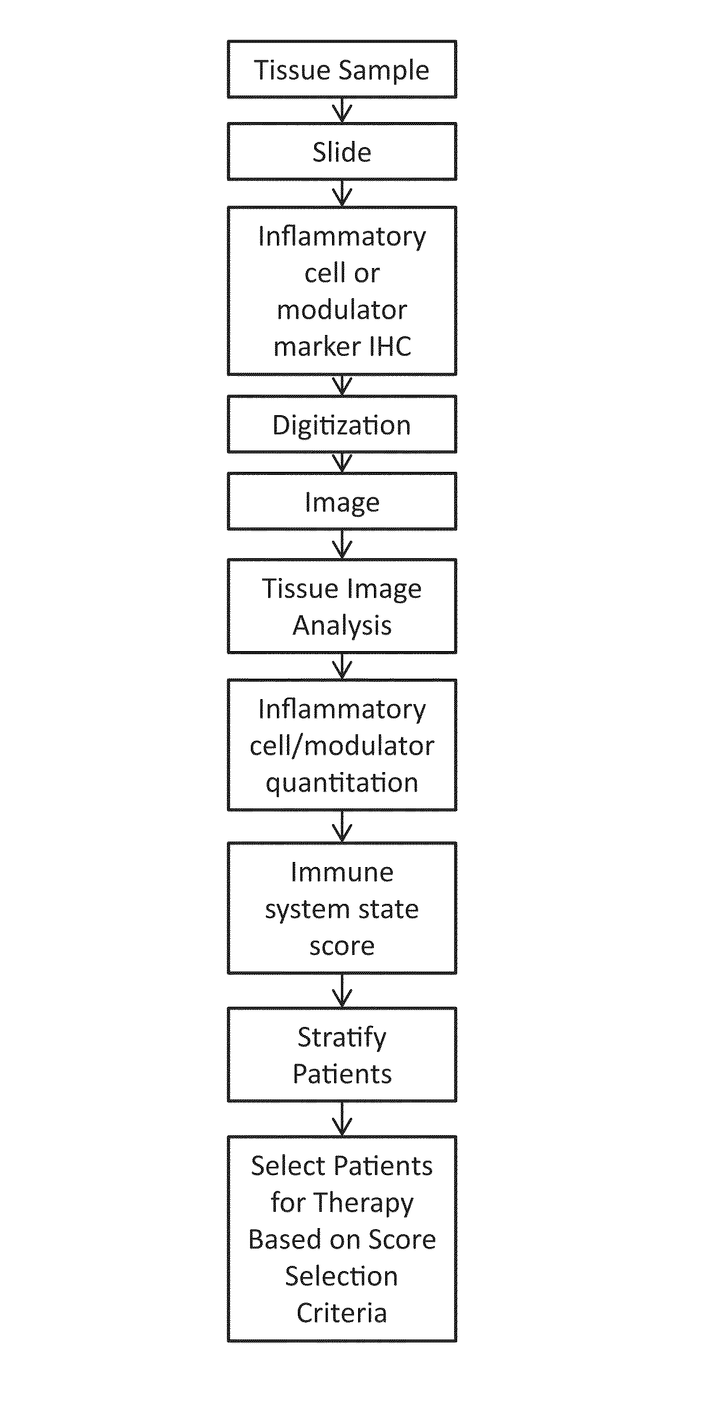
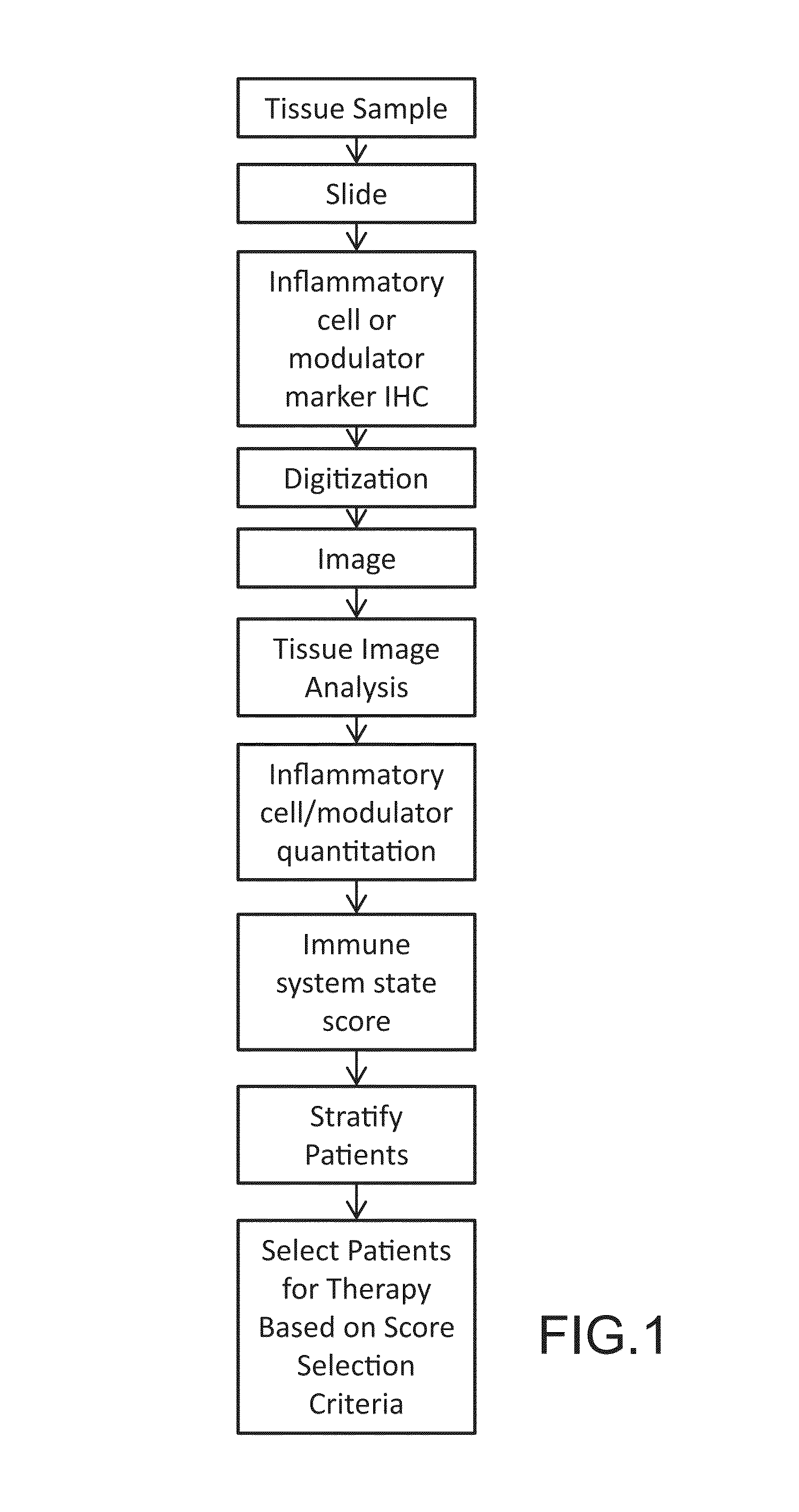
[0029]In an embodiment as illustrated in FIG. 1, a method analyzing inflammatory cells and mediators of inflammation in biological tissue sections containing cancerous lesions may generally comprise: (i) tissue acquisition, preparation, and embedding; (ii) slide preparation; (iii) immunohistochemical staining for cells, inflammatory cells, and / or modulators of the inflammatory response; (iv) digitization of the slide to produce an image; (v) tissue image analysis and extraction of cell-based features by a software system which are saved to a database; (vi) quantitat...


PUM


Abstract

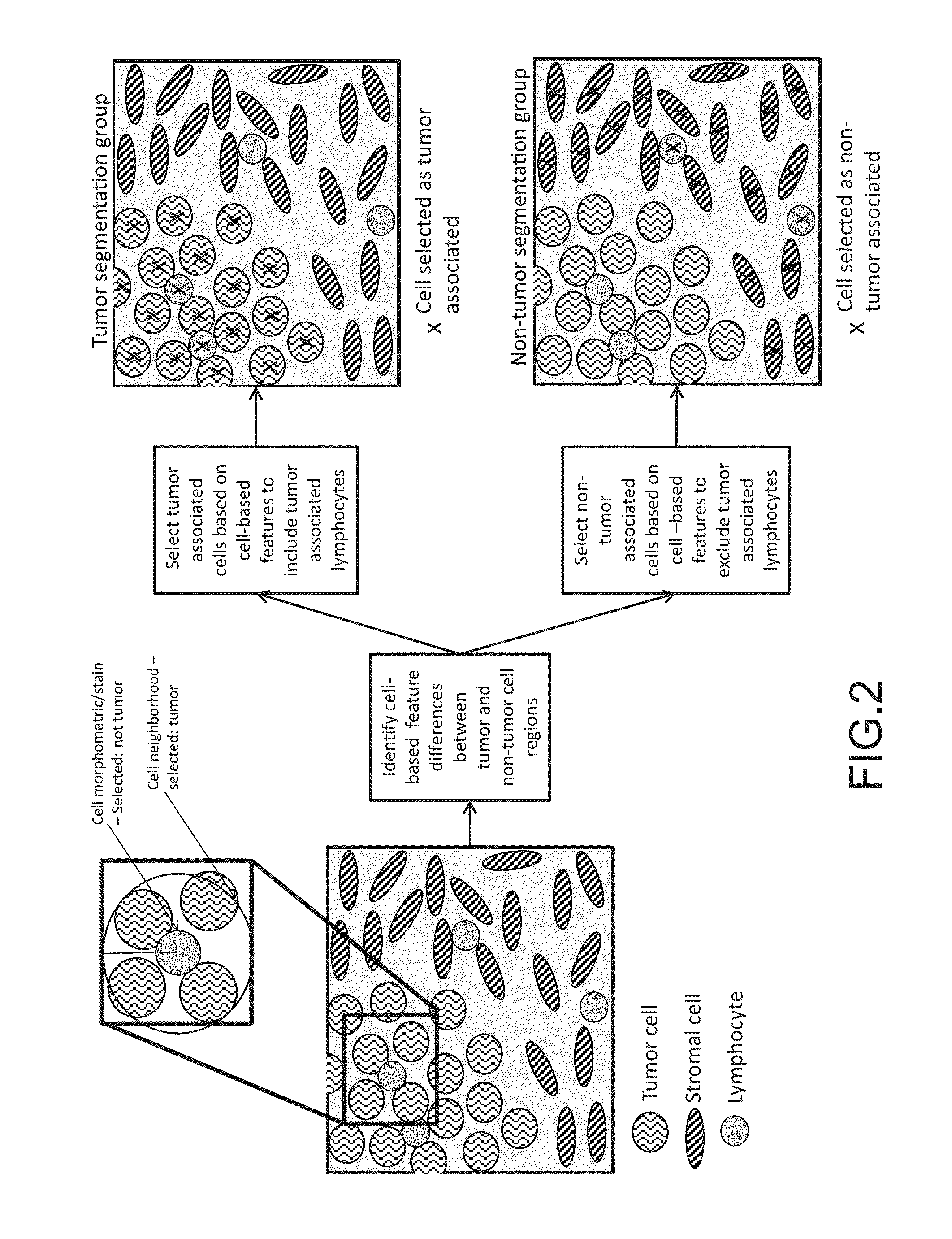
This disclosure concerns methods for evaluating inflammatory cells and modulators of the inflammatory response in tumor tissue and other relevant tissue types. The methods entail: obtaining a tissue sample and processing said tissue sample to produce histologic slides of tissue sections; staining of the tissue sections to identify inflammatory cells and modulators of the inflammatory response; digitizing slides to produce an image of the stained tissue sections; digitally stratifying the tissue sample into tumor and other relevant tissue compartments; and using digital image analysis to quantify cell-based and cell population-based features. The quantification of cell-based and cell population-based features within a tissue compartment of interest is used to develop a summary score of the immune system-tissue compartment of interest interaction. Patient stratification and selection as candidates for a therapeutic approach is ultimately based on the summary score value.

Description

CROSS-REFERENCE TO RELATED APPLICATIONS[0001]This application claims benefit of priority with U.S. Provisional Ser. No. 62 / 049,954, filed Sep. 12, 2014; the contents of which are hereby incorporated by reference.BACKGROUND[0002]1. Field of the Invention[0003]The disclosure relates to methods for quantitative assessment of a patient-specific immune system state in tumor tissue and other relevant tissue compartments for evaluating a potential or ongoing treatment; and more particularly to such methods including analyzing inflammatory cell quantity, expression level of biomarkers or quantity of mediators of inflammation, and the distribution pattern of inflammatory cells in distinct tissue compartments within tissue samples containing tumor tissue.[0004]2. Description of the Related Art[0005]The role of the immune system in response to cancer treatment is becoming more evident. Research studies have shown that the localization of specific inflammatory cell types in or near tumor tissue...

Claims


Application Information

Patent Timeline
IPC IPC(8): G06K9/00G06T7/00
CPCG06K9/00127G06T7/0012G06T2207/30004G06T2207/10024G06T2207/10056G06T2207/30024G06T2207/30096G06V20/69
Inventor PELJTO, MIRZAMILICI, ANTHONY, J.LANGE, HOLGERKRUEGER, JOSEPHAEFFNER, FAMKEYOUNG, GEORGE, D.MARTIN, NATHAN, T.
Owner FLAGSHIP BIOSCI
Learn More