Method of retrieving data from a medical image data set

a medical image and data retrieval technology, applied in the field of retrieving data from medical image data sets, can solve the problems of time-consuming, inconvenient, and inability to facilitate or accelerate the finding of target locations, and achieve the effect of improving the overview of a relatively large medical image data set and shortening user waiting times

Active Publication Date: 2016-09-06
KONINK PHILIPS ELECTRONICS NV
View PDF8 Cites 0 Cited by
  • Summary
  • Abstract
  • Description
  • Claims
  • Application Information

AI Technical Summary

Benefits of technology

[0012]Accordingly, the invention preferably seeks to mitigate, alleviate or eliminate one or more of the above mentioned disadvantages either singly or in any combination. In particular, it may be seen as an object of the present invention to provide a method of easier and / or faster retrieving of representative data from a medical image data set.
[0015]The invention is particularly, but not exclusively, advantageous for obtaining a method where a user may have an improved overview of a relatively large medical image data set. Additionally, the method may provide shorter user waiting times, because a user does not have to wait for a large data set to be retrieved since the representative indicator is typically only a fractional size of the data set. Also, the user does not have to scroll forward and backward through the acquired slices to locate the slices related to an area or region of interest (ROI).

Problems solved by technology

This is a quite time consuming task, which includes navigating to the areas of interest and visually inspecting the slices.
When inspecting the data like this, there is no help or assistance available to facilitate or accelerate finding of the target locations.
All applications that require retrieving data from large data sets, for inspection of objects that are represented by parts of those data sets, take time.
Loading large data sets takes even more time, which is annoying for the user.
If only a relatively small part of the data set is required, because the area of interest is much smaller that the complete covered volume, time and computer resources are wasted.
The problem of (nearly) useless data retrieval is becoming more and more pressing due to the increasing spatial resolution of current medical imaging modalities.
Thus, previously maybe around 100-200 slices were obtained from an imaging session, now the number can typically be above 1000 slices, making manual analysis of such an amount of slices quite time consuming.
However, the thumbnail pictures are chosen based on a numbering of the slices or pictures, which is not very effective for selecting relevant anatomical parts in the region of interest.

Method used


Image

  • Method of retrieving data from a medical image data set
  • Method of retrieving data from a medical image data set
  • Method of retrieving data from a medical image data set

Examples


Embodiment Construction

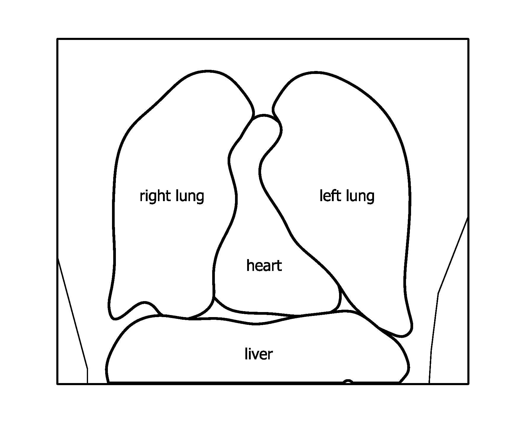
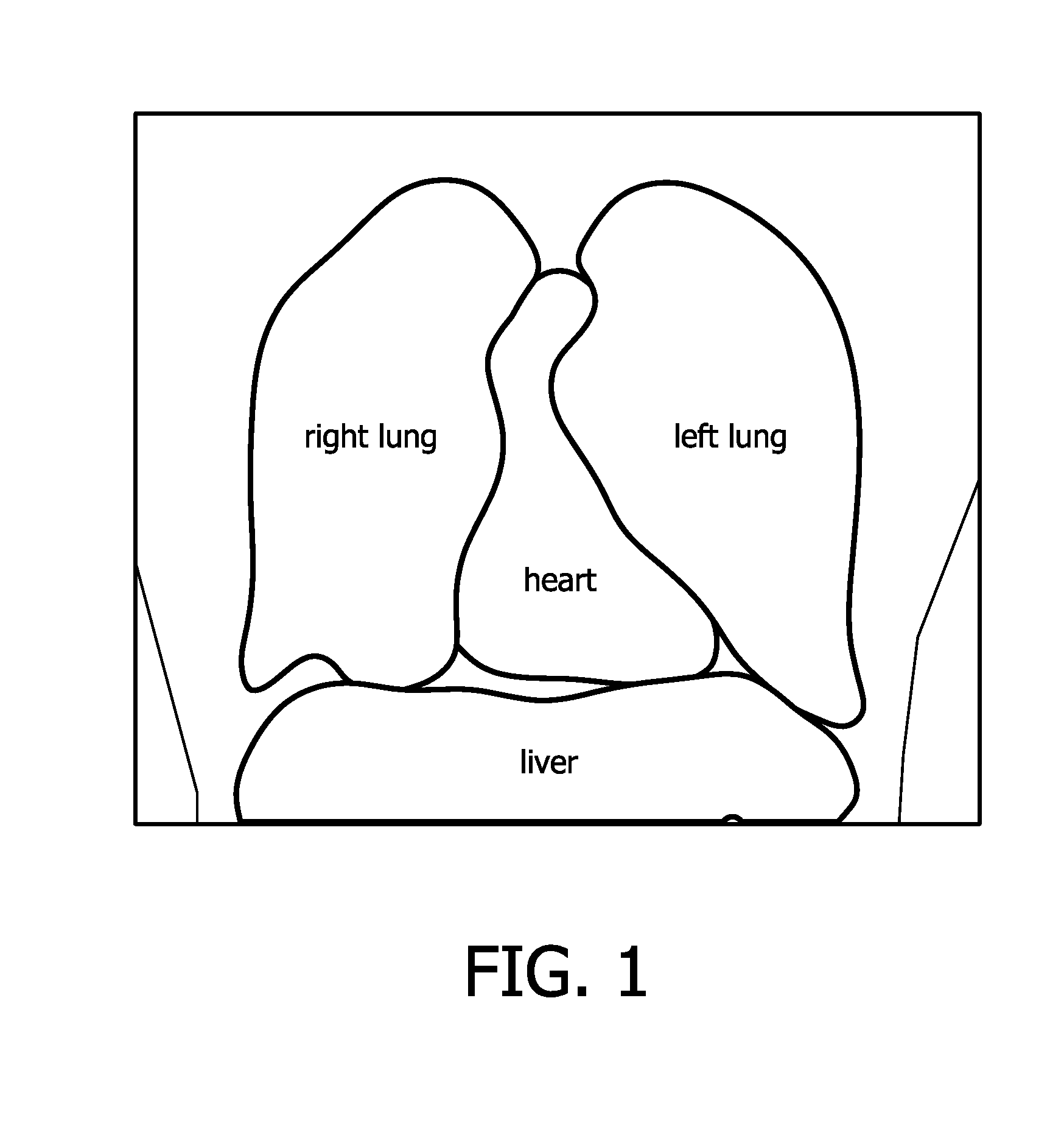
[0045]FIG. 1 shows a schematic drawing of various organs found from a content aware analysis of the upper abdomen and the thorax of a human being (homo sapiens). In currently available systems for performing a content specific analysis with a semi-automated / automated anatomical recognition method, it is possible to analyze and recognize organs like lungs (left and right), heart, and liver (all three organs shown in FIG. 1), but also spleen, spinal cord, brain, bladder, colon, vessel structures in general, and kidneys are presently detectable from a medical image data set 5. For general reference to segmentation in medical imaging, the reader is referred to C. Xu, D. Pham, and J. L. Prince, “Medical Image Segmentation Using Deformable Models,”Handbook of Medical Imaging, Volume 2: Medical Image Processing and Analysis, pp. 129-174, edited by J. M. Fitzpatrick and M. Sonka, SPIE Press, May 2000, which is hereby incorporated by reference in its entirety.

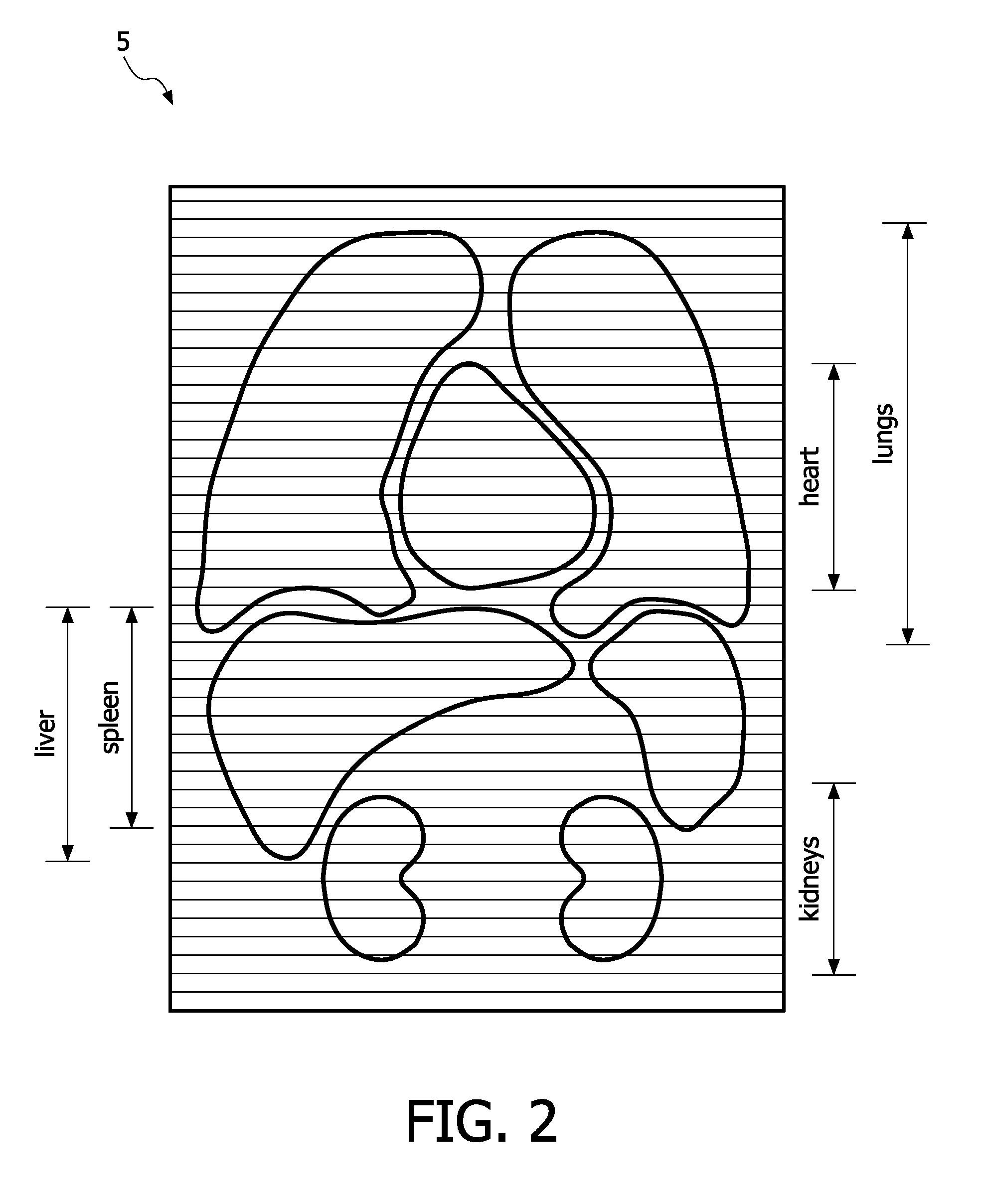
[0046]FIG. 2 shows a schematic d...


PUM


Abstract

The present invention discloses a method of retrieving a plurality of data slices from a medical image data set (5), the method comprising the steps of: a) displaying an indicator (10, 20) associated with the plurality of data slices; b) selecting the indicator (10, 20) based on a user input; and c) retrieving the plurality of data slices (1, 2) associated with the indicator when said indicator is selected; wherein the association between the indicator and the plurality of slices is based on segmentation of the medical image data set, the indicator representing an object obtained in the segmentation of the medical image data set, the plurality of data slices comprising the object data. The method of the invention reduces the amount of data transfer because it allows for retrieving only those data slices which comprise relevant data relating to the object of interest.

Description

FIELD OF THE INVENTION[0001]The present invention relates to a method of retrieving data from a medical image data set obtained from a medical imaging modality. The invention also relates to a corresponding system for implementing the method, and a corresponding computer program product for such an implementation.BACKGROUND OF THE INVENTION[0002]3D medical image data is often acquired in the form of a set of slices of data. The set of slices defines volumetric, i.e. 3D, image data. This set may be referred to as an image data set or just as image data. The most common way of inspecting a large 3D medical data set from e.g. a CT scanner or similar device, is to load a data set from a storage medium and to display cross-sectional slices of that volume. The locations of interest in that volume are found by scrolling through the (often large number of) slices. Normally, this is the stack of original slices as they come from the acquisition device. This is a quite time consuming task, wh...

Claims


Application Information

Patent Timeline
Patent Type & Authority Patents(United States)
IPC IPC(8): G06K9/00G06F19/00G16H10/60G16H30/20G16H30/40
CPCG06F19/321G16H30/20G16H30/40
Inventor LOBREGT, STEVENFREDERIK, JOOSTTJHANG, ALAN PEK SENG
Owner KONINK PHILIPS ELECTRONICS NV
Why Eureka
Try Eureka