Methods and systems for prognosis and diagnosis of brain damage

Active Publication Date: 2016-11-01
UNIV OF KENTUCKY RES FOUND
View PDF6 Cites 4 Cited by
  • Summary
  • Abstract
  • Description
  • Claims
  • Application Information

AI Technical Summary

Benefits of technology

The patent text discusses the need for a biomarker that can be used to diagnose and prognosis brain damage, particularly in the field (e.g. in the case of traumatic brain injury or stroke). Current methods for diagnosis and treatment are limited, and there is a need for a biomarker that can be measured quickly and accurately to improve the diagnosis and treatment of brain damage.

Problems solved by technology

Similarly, about 795,000 Americans suffer a stroke each year, and stoke is a leading cause of serious long-term disability.
Current diagnostic and prognostic tools used for evaluation of TBI and stroke remain limited particularly for in field (i.e., pre-hospital) evaluation.
Although CT is sensitive in the identification of acute hemorrhage and fracture associated with severe injury, it is relatively insensitive in the identification of diffuse injury associated with milder TBI.
MRI is capable of detecting hematomas, small contusions, and ischemic events not captured by CT, but it is relatively insensitive to diffuse axonal injury and may not be feasible in all situations.
Furthermore, the time delay associated with current diagnostic approaches for stroke patients limits the time during which thrombolytics (tissue plasminogen activator; t-PA) may be administered.
Because of the potential detrimental effects of use oft-PA in patients with a small hemorrhagic stroke, the lack of a diagnostic that can quickly differentiate between hemorrhagic and ischemic stroke is associated with underuse of fibrinolytic treatment.
Therefore, mild TBI is difficult to diagnose because mild TBI can present limited injury and only subtle changes in GCS scores.
However, known biomarker have limited capabilities For instance, even though known biomarkers may identify subjects having experienced a stroke from normal subjects, known biomarkers are not capable of delineating between ischemic stroke patients and patients having experienced a hemorrhagic stroke, seizure, migraine, syncope, hypoglycemia, and the like.
Furthermore, although some biomarkers have been identified as potentially being useful for characterizing brain damage, most known biomarkers are quantified using time-consuming techniques, such as enzyme linked immunoassays (ELISAs) that can take as long as 24 hours to complete.
Therefore, known techniques provide limited information on a subject only after an optimal time period for treatment has passed.

Method used


Image

  • Methods and systems for prognosis and diagnosis of brain damage
  • Methods and systems for prognosis and diagnosis of brain damage
  • Methods and systems for prognosis and diagnosis of brain damage

Examples


example 1

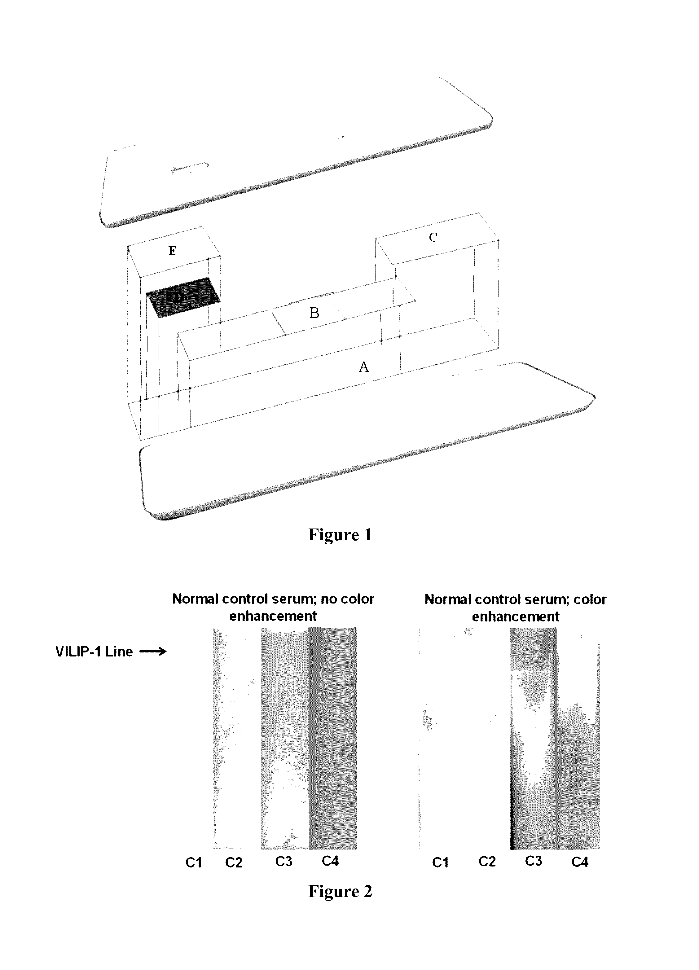
[0067]This Example describes a method for preparing a lateral flow device for detecting the presence of VILIP-1 in a biological sample. As shown in FIG. 1, the lateral flow device comprises of a combination of membranes that remove cells (e.g., whole blood) and serum albumin (e.g., rat or human) from a biological sample, nanogold labeled anti VILIP-1 raised against the N terminus of human VILIP-1, and a narrow line near the terminal end of anti-VILIP-1 antibody raised against the C terminus of human VILIP-1. More specifically, and still referring to FIG. 1, the lateral flow device comprised a laminate backing (A; MIBA-010; Diagnostic Consulting Network, Carlsbad, Calif.), a sample portion for receiving a biological sample (E; Vivid Plasma Separation-GX; Pall Corporation, Port Washington, N.Y.), a conjugate portion (D; Pall, Grade 896), a detection portion (B; Unisart CN140 Nitrocellulose Membrane, 42 mm; Sartorius Stedin North America Inc, Bohemia, N.Y.), and a wicking portion (C; W...

example 2

[0073]This Example describes procedures conducted to evaluate how the embodiments of lateral flow devices that were prepared in Example 1 can be used to detect the presence of VILIP-1 in subjects having brain damage, including brain damage caused by ischemic stroke or TBI.

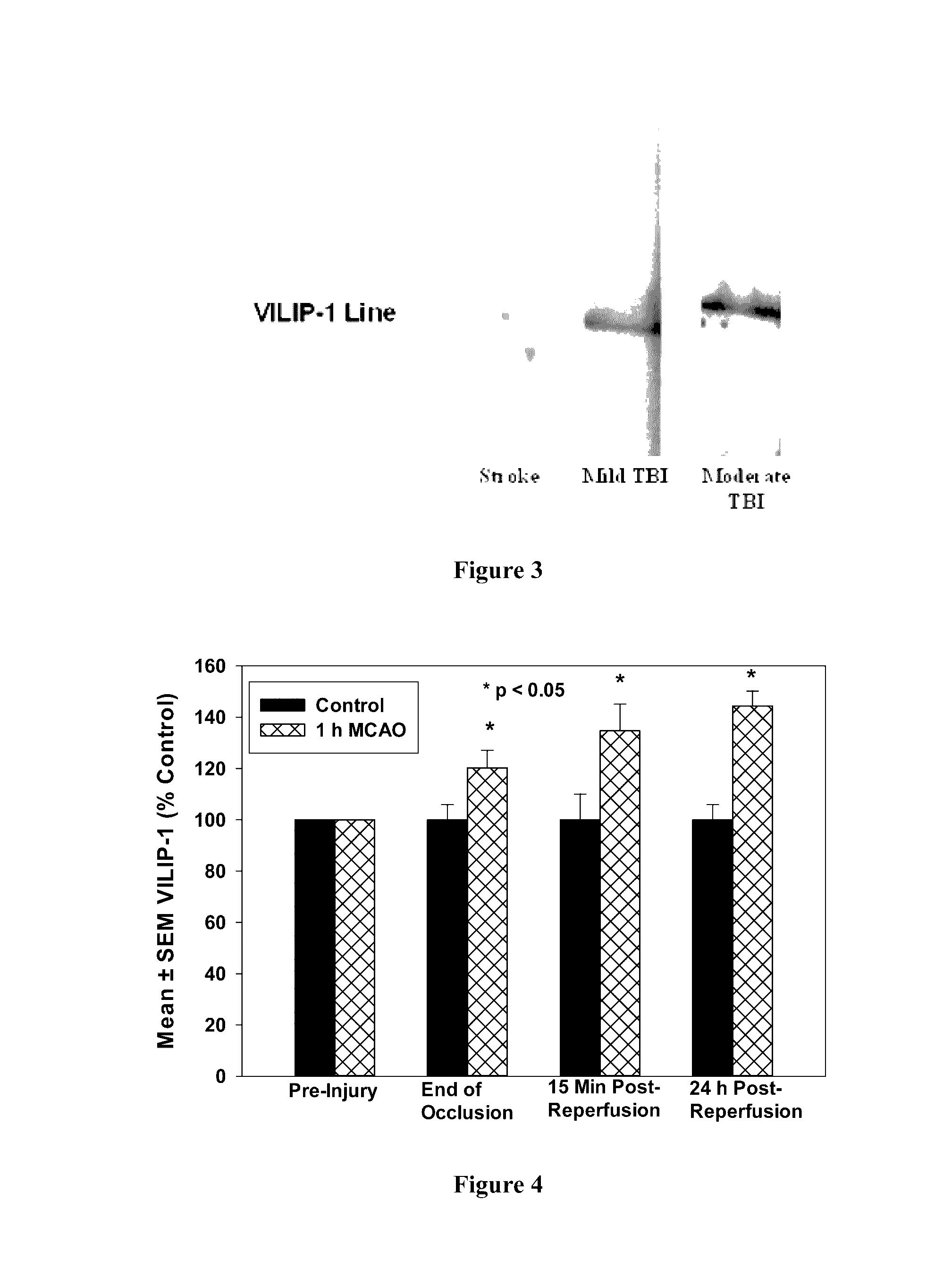
[0074]First, using recombinant VILIP-1 as a standard protein, a limit of detection of 0.2 ng / ml serum and a linear response between 0.2 ng / ml and 7.0 ng / ml was detected (r2=0.91). FIG. 2 shows levels of VILIP-1 in normal control serum are below the limit of detection. Thus, the lateral flow devices were capable of detecting the presence of VILIP-1 in biological samples.

[0075]To determine if serum VILIP-1 levels change in due to ischemic stroke, animals were subjected to a 1 hour middle cerebral artery occlusion (MCAO) using the Zea Longa method. Specifically, nine three-month-old Sprague-Dawley rats (250-300 g) were subjected to a 1 hour ischemic event induced by suture occlusion of the middle cerebra artery. Contr...

example 3

[0078]This Example describes the relationship between VILIP-1 levels and brain damage that were observed using the procedures described in Example 2. Specifically, this Example describes how VILIP-1 levels were affected by the induced ischemic stroke and TBI in rat subjects.

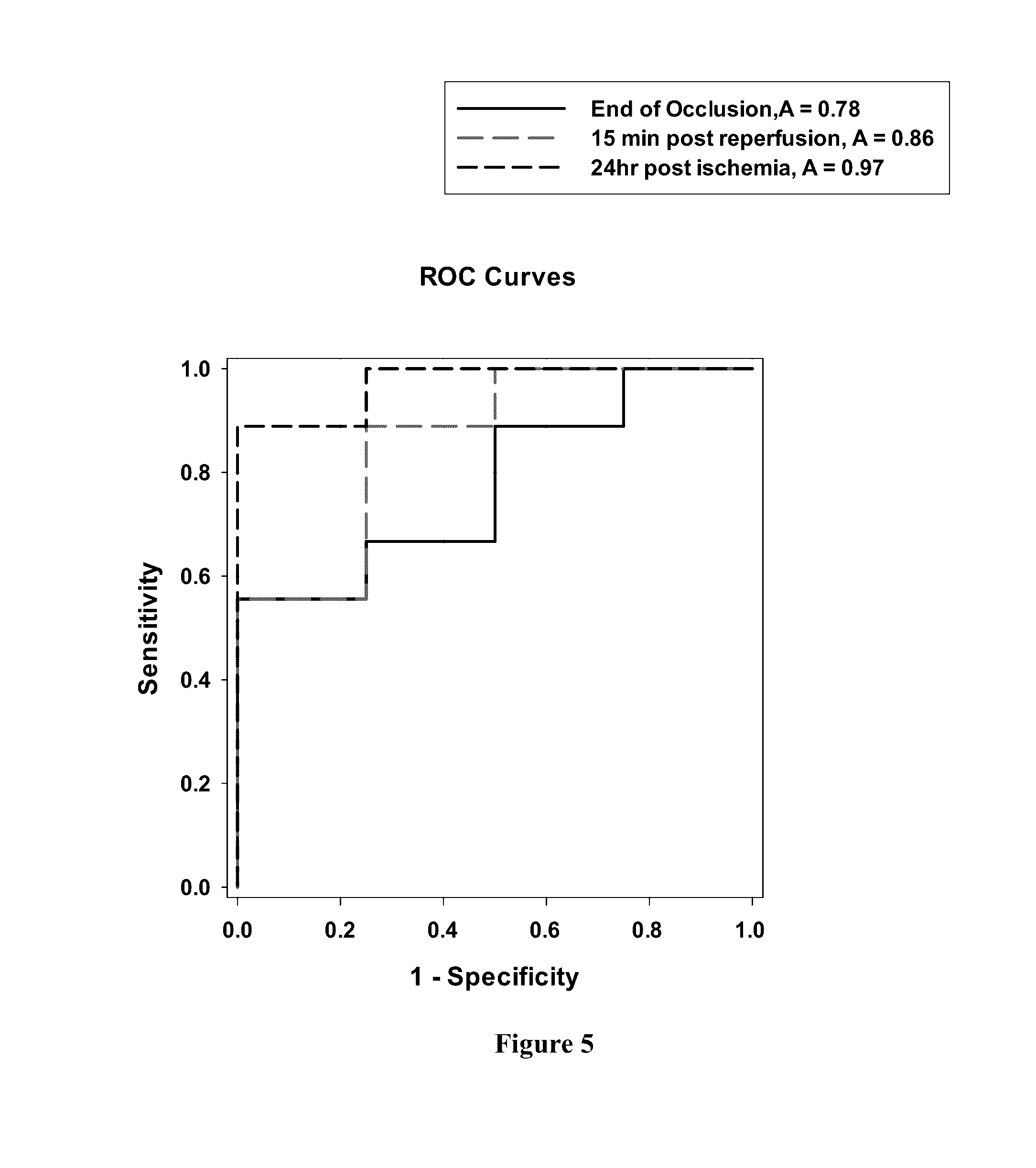
[0079]As described above, whole blood (10 μL) were analyzed at the time of draw and the remaining blood samples were centrifuged to prepare serum which was analyzed from all time points later. FIG. 3 shows that levels of VILIP-1 in serum were considerably higher in mild TBI compared to ischemic stroke 1 hour and 15 minutes post injury, thereby suggesting that TBI can induce more pronounced neuronal injury (i.e., brain damage). Without being bound by theory or mechanism, VILIP-1 levels for subjects having experienced hemorrhagic stroke can be comparable to those observed in TBI because hemorrhagic stroke can cause severe tissue damage. Thus, optionally when combined with clinical presentation, the presence of VILI...


PUM


Abstract

The presently-disclosed subject matter includes methods and devices for diagnosing, prognosing, and treating brain damage in a subject, including brain damage caused by stroke or a traumatic brain injury (TBI). The methods can comprise providing a sample obtained from the subject, exposing the sample to an antibody selective for a visinin-like protein, detecting the presence of a complex that includes the antibody and the visinin-like protein, and diagnosing and / or prognosing the subject as having brain damage if there is the presence of the complex. Embodied methods can also comprise administering a treatment for brain damage if the subject includes the presence of the visinin-like protein.

Description

RELATED APPLICATIONS[0001]This application claims priority from U.S. Provisional Patent Application No. 61 / 860,645, filed Jul. 31, 2013, the entire disclosure of which is incorporated herein by this reference.TECHNICAL FIELD[0002]The presently-disclosed subject matter relates to methods and systems for prognosis, diagnosis, and treatment of brain damage in a subject. In particular, the presently-disclosed subject matter relates to methods and systems for prognosing, diagnosing, and treating brain damage that include measuring levels of visinin-like family proteins in a biological sample obtained from a subject.INTRODUCTION[0003]Traumatic brain injury (TBI) is a significant civilian and military hearth concern. Brain injury can exist in individuals who experience concussive or sub-concussive blows to the head, and has particular relevance to athletic and military activities. Without reliable diagnostics that can determine whether a blow to the head affected the brain, athletes and so...

Claims


Application Information

Patent Timeline
IPC IPC(8): G01N33/68
CPCG01N33/6872G01N33/6893G01N2800/2871G01N2333/4727G01N33/558G01N33/54388
Inventor LOVELL, MARK A.LYNN, BERT C.BRADLEY-WHITMAN, MELISSA A.
Owner UNIV OF KENTUCKY RES FOUND
Why Eureka
Try Eureka