
Modulating gantry rotation speed and image acquisition in respiratory correlated (4D) cone beam CT images

a technology of gantry rotation speed and image acquisition, which is applied in the field of radiotherapy, can solve the problems of blurry image reconstruction, increased the likelihood that some of the radiation targeted at the tumor will irradiate healthy lung tissue, and slow image reconstruction process, so as to minimize the time required, minimize the angular separation, and maximize the effect of image quality

Active Publication Date: 2017-01-10
THE BOARD OF TRUSTEES OF THE LELAND STANFORD JUNIOR UNIV +1
View PDF9 Cites 9 Cited by
  • Summary
  • Abstract
  • Description
  • Claims
  • Application Information

AI Technical Summary

Benefits of technology

This approach reduces the radiation dose to the patient, enhances image quality, and allows for real-time adaptation to irregular breathing patterns, improving the angular separation of projections and the likelihood of acquiring suitable quality images during 4DCBCT imaging.

Problems solved by technology

However, imaging techniques such as MRI, PET, CT and CBCT are blurry or contain artifacts when there is significant respiratory motion.
As a consequence, it is difficult for a radiotherapist to accurately position a lung tumor patient for treatment, which increases the likelihood that some of the radiation that is targeted at the tumor will irradiate healthy lung tissue.
Image reconstruction can be a slow process that can take several minutes to reconstruct a single image.
The resulting image is likely to contain artifacts, or the image will be blurry, in the region where significant respiratory motion takes place.
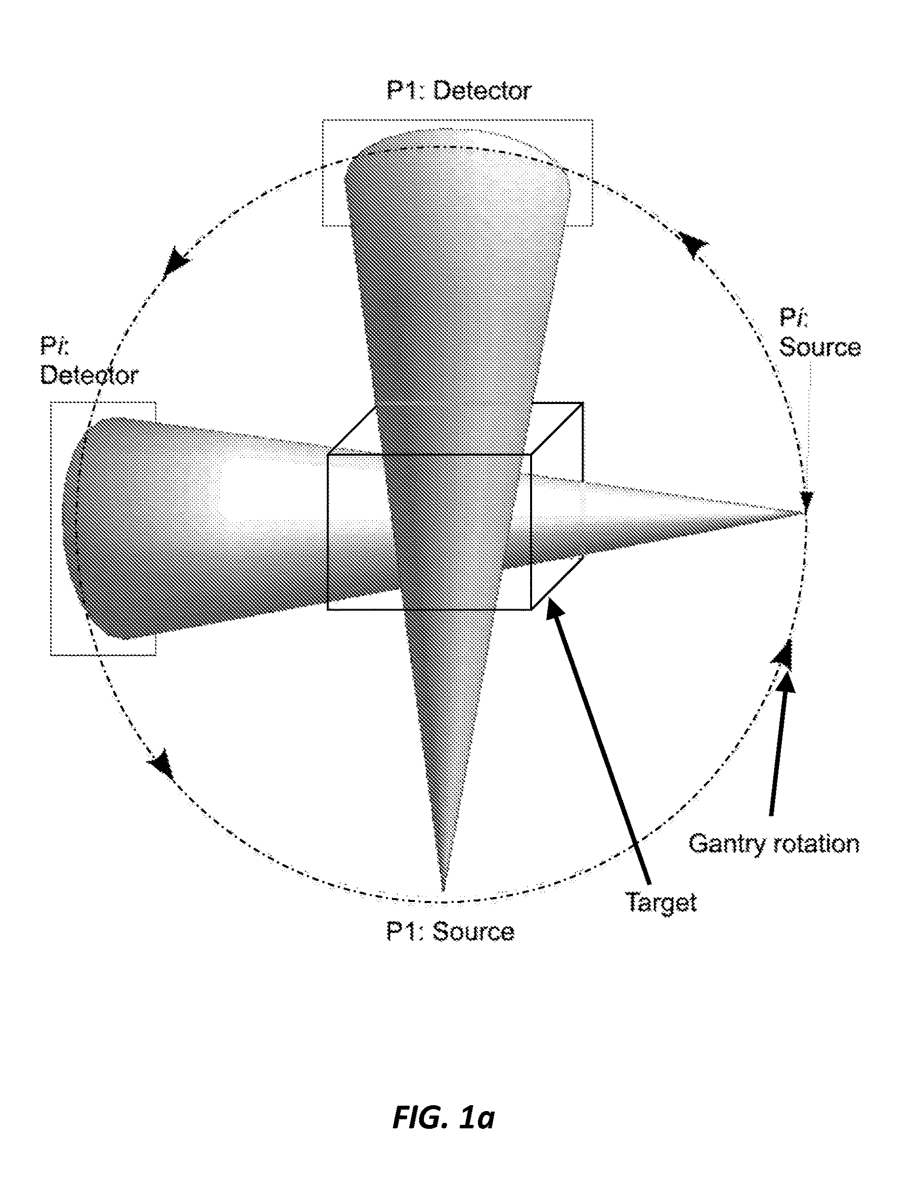
However, as can be seen in the polar plot in FIG. 2c, if the gantry is rotated around the patient with a constant angular velocity the angular separation between projections is not ideal and projection clustering occurs.
These methods assume that the patient's breathing is regular so the algorithms are likely to have difficulty reconstructing an image if the patient's breathing is irregular.
However, in the current implementation there is no communication between the respiratory signal and image acquisition.
This results in bunched angular projections, with the CBCT images suffering from poor image quality and streak artifacts.
The poor quality limits the use of current images for online guidance and anatomic and functional adaptation.
However, the use of a constant angular velocity results in clustering of projections.
The patient has received a radiation dose from 60 projections but there are not enough projections to reconstruct an image of suitable quality.
In practice the displacement bin can be missed during both inhale and exhale in consecutive cycles leading to large gaps between projections.
Because projections are post-processed into respiratory bins, first generation methods are unable to adapt if the patient's breathing is irregular as no feedback with the patient's respiratory data is used to regulate the gantry.
Therefore image quality is likely to be poor if the patient's breathing is irregular.

Method used


Image

  • Modulating gantry rotation speed and image acquisition in respiratory correlated (4D) cone beam CT images
  • Modulating gantry rotation speed and image acquisition in respiratory correlated (4D) cone beam CT images
  • Modulating gantry rotation speed and image acquisition in respiratory correlated (4D) cone beam CT images

Examples


Embodiment Construction

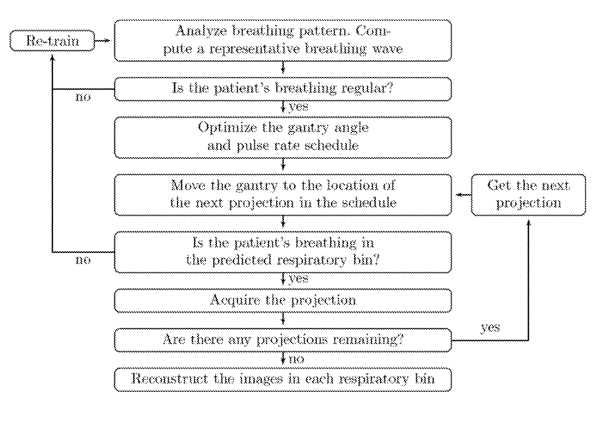
[0044]Four Dimensional Cone Beam Computed Tomography (4DCBCT) images of lungs are rarely used by radiotherapists to position a patient for treatment because of blurring as a result of respiratory motion. One embodiment of the current invention provides a novel Mixed Integer Quadratic Programming (MIQP) model for Respiratory Motion Guided-4DCBCT (RMG-4DCBCT), which can be used to obtain 4DCBCT images of the lungs and lung tumors by a radiotherapist in the treatment room prior to treatment. RMG-4DCBCT regulates the gantry angle and projection pulse rate, in response to the patient's respiratory signal, so that a full set of evenly spaced projections can be taken in a number of phase, or displacement, bins during the respiratory cycle. In each respiratory bin, an image is reconstructed from the projections to give a 4D view of the patient's anatomy so that the motion of the lungs, and tumor, can be observed during the breathing cycle. A solution to the full MIQP model in a practical am...


PUM


Abstract

A method of optimizing 4D cone beam computed tomography (4DCBCT) imaging is provided that includes using a scanner to generate projections of a target, where the projections are used to form a cone beam computed tomography (CBCT) scan of the target, where the CBCT includes a 3D image of the target, and using an appropriately programmed computer to control rotation speed of a gantry and projection acquisition of the CBCT in real-time according to a measured patient respiratory signal, where the real-time acquisition of the 4CBCT forms an optimized 4DCBCT image set.

Description

CROSS-REFERENCE TO RELATED APPLICATIONS[0001]This application is a 371 of PCT / US2012 / 048693 filed on Jul. 27, 2012. PCT / US2012 / 048693 filed on Jul. 27, 2012 claims the benefit of US 61 / 574126 filed on Jul. 28, 2011 and US 61 / 671028 filed on Jul. 12, 2012.STATEMENT OF GOVERNMENT SPONSORED SUPPORT[0002]This invention was made with Government support under contract CA116602 awarded by the National Institutes of Health. The Government has certain rights in this invention.FIELD OF THE INVENTION[0003]The present invention relates generally to radiotherapy. More particularly, the invention relates to obtaining 4DCBCT images of the lungs and lung tumors using mathematical optimization models for Respiratory Motion Guided-4DCBCT (RMG-4DCBCT).BACKGROUND OF THE INVENTION[0004]Lung cancer is the leading cause of cancer related death worldwide (18%) with 1.2 million new cases reported annually. Despite 36% to 71% of lung cancer patients receiving radiotherapy, and continuous efforts to improve t...

Claims


Application Information

Patent Timeline
Patent Type & Authority Patents(United States)
IPC IPC(8): A61B6/03A61B6/00A61N5/10
CPCA61B6/541A61B6/4085A61B6/486A61B6/5205A61B6/5264A61B6/583A61N2005/1061
Inventor KEALL, PAUL J.O'BRIEN, RICKYCOOPER, BENJAMIN J.LU, JUNWILLIAMSON, JEFFREY F.
Owner THE BOARD OF TRUSTEES OF THE LELAND STANFORD JUNIOR UNIV
Learn More