
System and method for projection image tracking of tumors during radiotherapy

a tumor and imaging system technology, applied in the field of system and method for projection image tracking of tumors during radiotherapy, can solve the problems of destroying tumors, and reducing the accuracy and effectiveness of radiotherapy treatment, so as to improve the efficiency of tumor treatment, reduce the distribution of photon energy, and high contrast imaging

Inactive Publication Date: 2017-01-17
PARTAIN LARRY D
View PDF39 Cites 2 Cited by
  • Summary
  • Abstract
  • Description
  • Claims
  • Application Information

AI Technical Summary

Benefits of technology

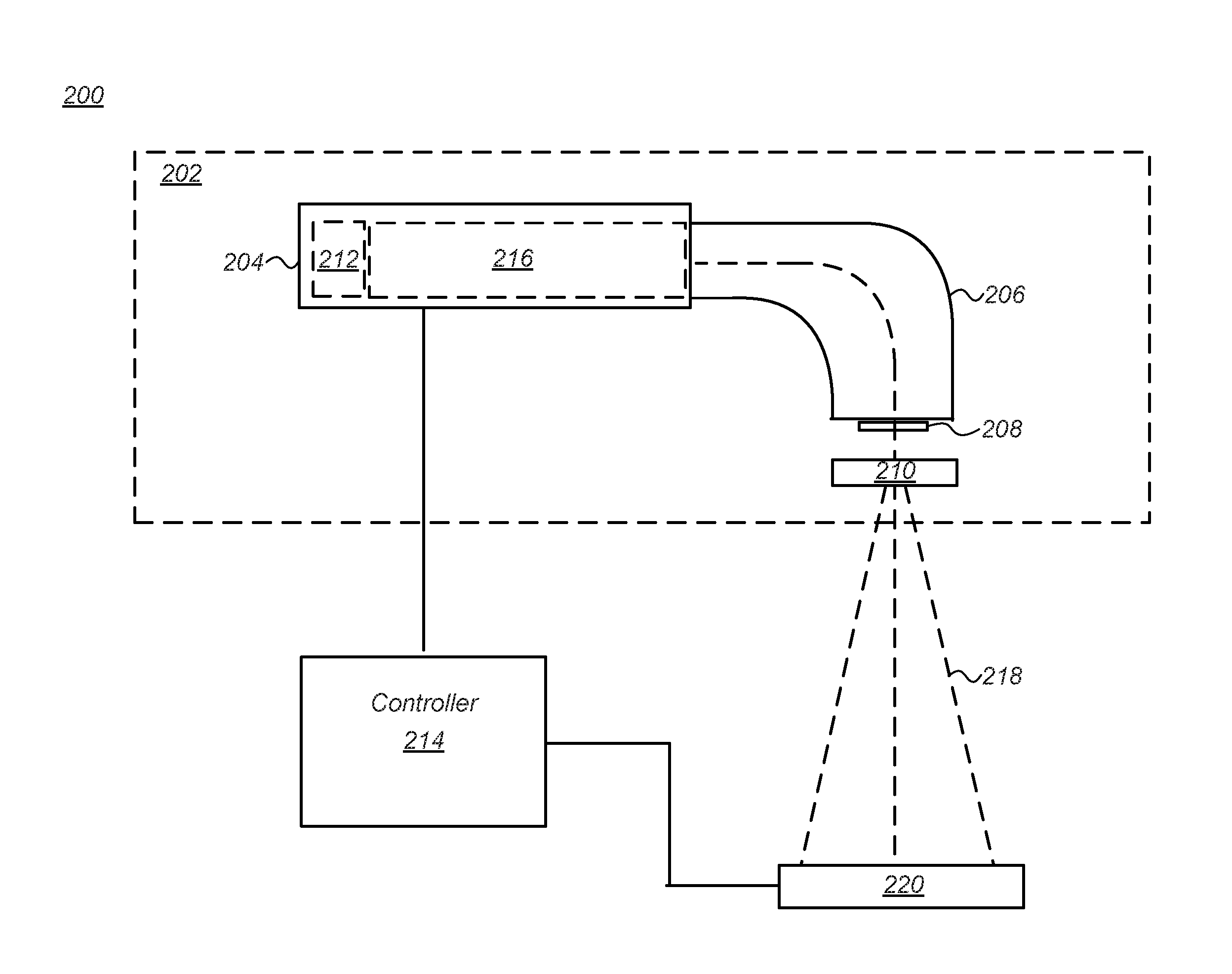
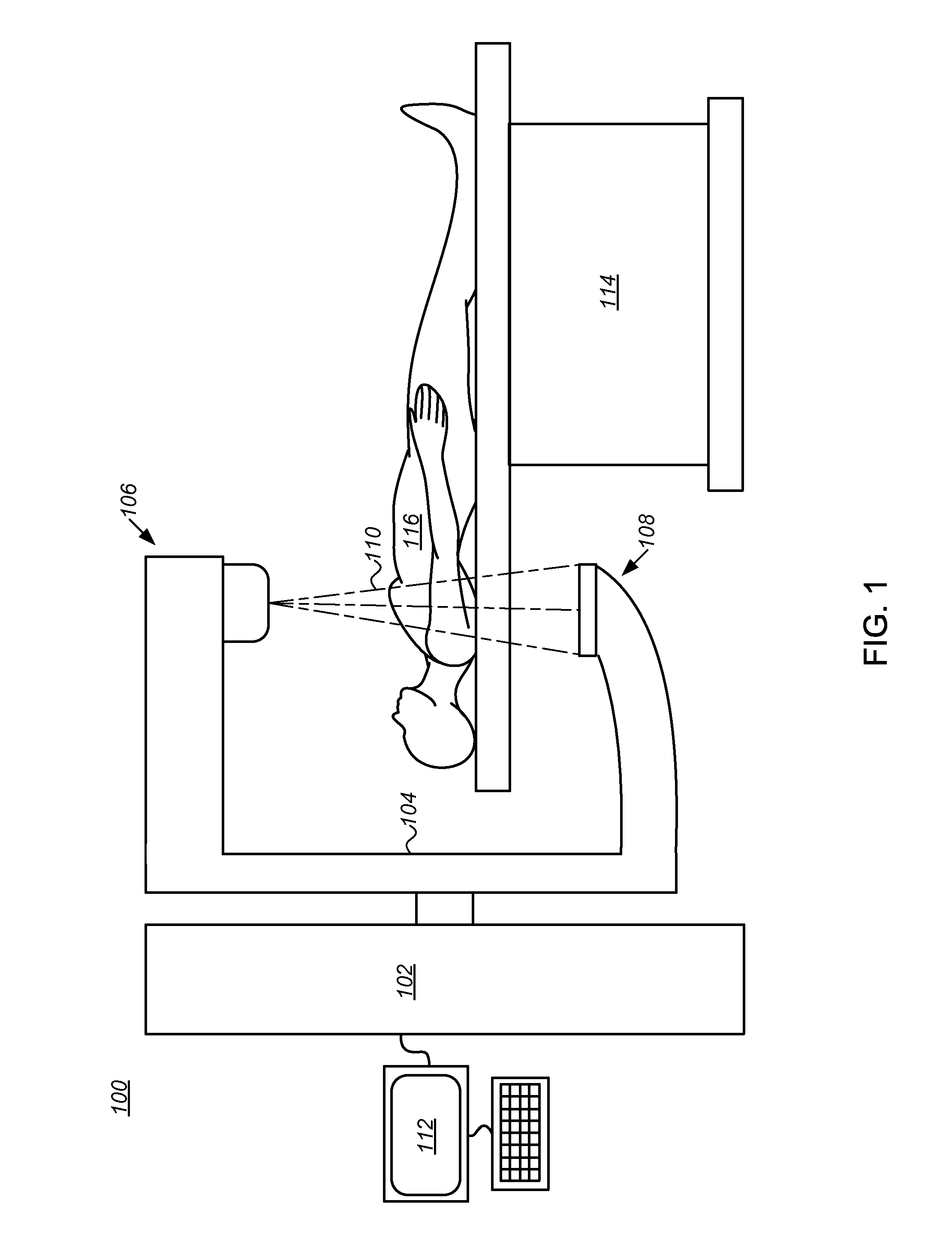
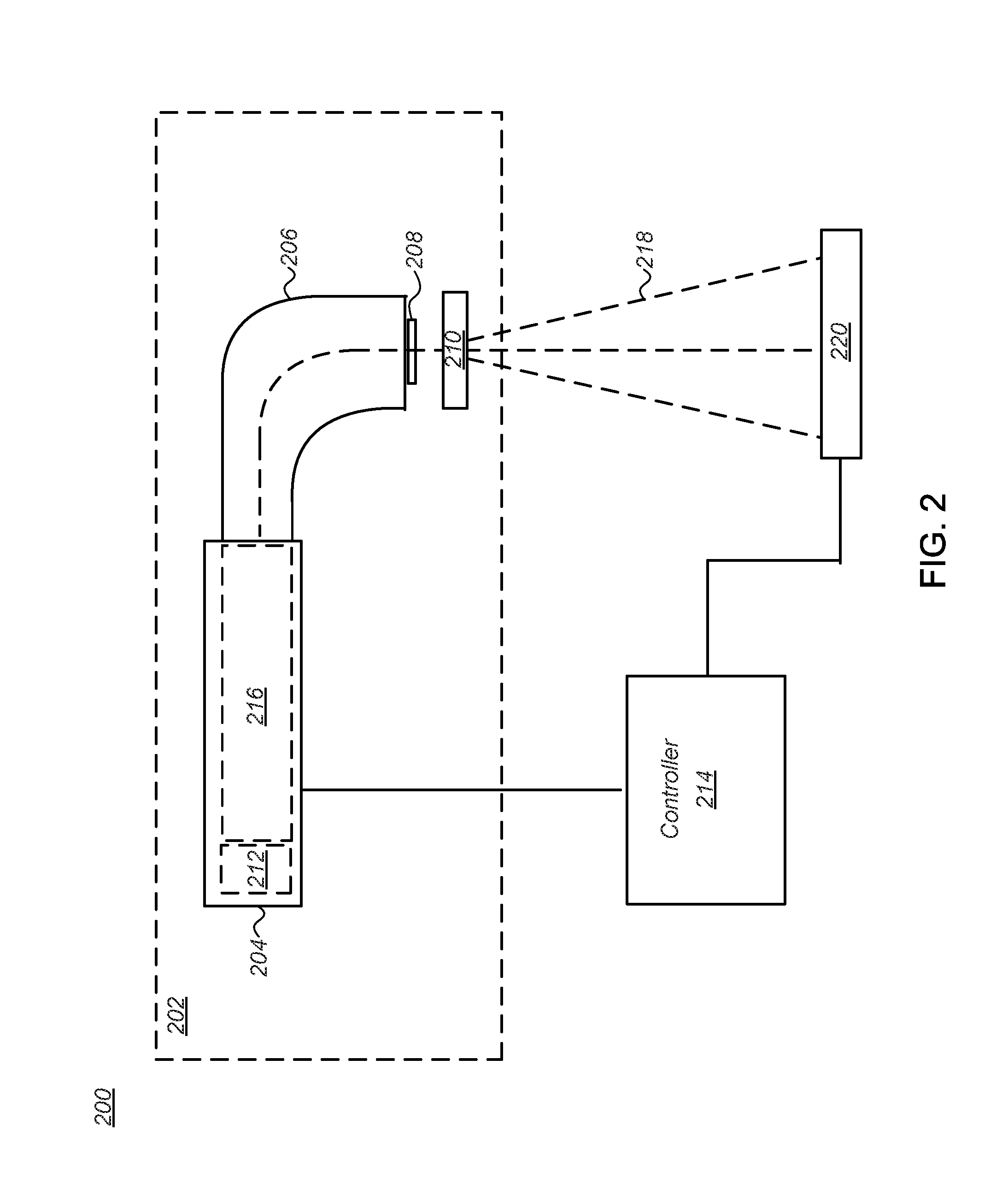
The invention is a system and method for imaging and treating tumorous tissue in a patient using a radiation source. The system generates a beam of high-energy photons that can be used for both contrast imaging and treatment. The system includes a charged particle accelerator, a target, a collimator, and an imaging device with high efficiency. The method involves applying charged particles to a target to generate the radiation beam, defining a target region of tumorous tissue, determining the location and dose for treatment, and controlling the radiation beam to deliver it to the target region. The technical effects include improved imaging and treatment of tumorous tissue using a single system and method.

Problems solved by technology

The ionizing radiation damages the DNA of tissue in the tumor leading to cellular death and destroying the tumor.
This can be problematic as in many therapies using conventional radiation therapy systems, since the patient's anatomy will often shift or move during the treatment, for example breathing induced movement.
This movement can significantly reduce the accuracy and effectiveness of the radiotherapy treatment, particularly for therapies such as hypofractionated stereotactic body radiotherapy (SBRT) in which a highly collimated beam is used to deliver a high biological dose of radiation directly to the tumor.
While useful for initial set-up, this approach has little utility in maintaining the collimated beam on the targeted tumor when the tumor moves unseen inside the patient.
However, in practice prior art EPID images exhibit low contrast.
This surrounding dose damage issue is a major and poorly solved problem currently in the field of radiation therapy.
In contrast the low DQE value for the conventional imager results in poor or low contrast images of the tumorous tissue making targeting of the tumor problematic, and can result in too high a dose being delivered to the patient when the system is operated in therapeutic mode and a signal from the imager is used to monitor dose.
As with the embodiment described with respect to FIG. 3 above, this lower DQE value for the conventional single layer imager results in low contrast images of the tumorous tissue making targeting of the tumor problematic, and can result in too high a dose being delivered to the patient.

Method used


Image

  • System and method for projection image tracking of tumors during radiotherapy
  • System and method for projection image tracking of tumors during radiotherapy
  • System and method for projection image tracking of tumors during radiotherapy

Examples


Embodiment Construction

[0018]The present disclosure is directed generally to projection image tracking of tumors during radiotherapy, and more particularly to a system and method for imaging and treatment of tumorous tissue in a patient.

[0019]The best cancer treatment delivery is obtained at MV (megavoltage) energies. It is possible to also image at MV energies and this can provide a tremendous targeting advantage because one can simultaneous see where the treatment dose is being delivered while the treatment process is in progress. The great advantage this gives is the opportunity to modulate in real time, the shape and dose distribution (across the shape) for maximum treatment dose delivery effectiveness to the cancer lesion while sparing surrounding healthy tissue from the strongly damaging effects of the radiation. This surrounding dose damage issue is a major and poorly solved problem currently in the field of radiation therapy.

[0020]Disclosed herein is a system and method that combines a reduction i...


PUM


Abstract

A system and method are provided for imaging and treatment of tumorous tissue in a patient. In an embodiment, the system includes a radiation source for generating a radiation beam comprising high-energy photons with low energy distributions for high contrast imaging along with high energy distributions for efficient treatment dose delivery. The radiation source includes a charged particle accelerator that generates charged particles having energies of less than 6 megavolts (MV), a target to receive the charged particles and generate the high-energy photons of the radiation beam, and a collimator to emit the radiation beam. The system further includes an imaging device of high detective quantum efficiency to define a target region of the tumorous tissue in the patient using the radiation beam, and a controller to determine the shape and modulate the dose for treatment of the tumorous tissue based on the defined target region, using the radiation beam.

Description

TECHNICAL FIELD[0001]The present disclosure relates generally to a system and method of providing radiation imaging and / or treatment, and more particularly to systems and methods for tracking of tumors during for projection image tracking of tumors during radiotherapy.BACKGROUND[0002]A Radiation therapy is widely used to identify and treat tumors in a patient. In radiation therapy, a high energy collimated beam of ionizing radiation is used to target tumorous tissue in the patient. The ionizing radiation damages the DNA of tissue in the tumor leading to cellular death and destroying the tumor. It will be understood that both the dose and placement of the ionizing radiation must be accurately controlled to ensure that the tumor receives sufficient radiation to destroy tumor tissue while damage to the surrounding healthy tissue is minimized. This can be problematic as in many therapies using conventional radiation therapy systems, since the patient's anatomy will often shift or move d...

Claims


Application Information

Patent Timeline
IPC IPC(8): A61N5/10
CPCA61N5/1067A61N5/1071A61N5/1064A61N5/1048A61N5/1049A61N5/1065A61N5/10A61N2005/1074A61N2005/1054
Inventor PARTAIN, LARRY D.
Owner PARTAIN LARRY D
Learn More