
Whole blood analytic device and method therefor

a technology of analytic devices and whole blood, which is applied in the direction of membrane technology, membranes, instruments, etc., can solve the problems of multiple steps, high cost of centrifuges, and difficult centrifugation

Active Publication Date: 2017-05-30
QUALIGEN INC
View PDF31 Cites 1 Cited by
  • Summary
  • Abstract
  • Description
  • Claims
  • Application Information

AI Technical Summary

Problems solved by technology

However, some difficulties exist with centrifugation.
For example, centrifuges are expensive, require the need for multiple steps (e.g., aliquoting through the use of precision pipette which can vary depending upon the technique of the operator), and are often unavailable at points of care such as a home, school, or bedside.
However, significant amounts of plasma may be retained and lost in the filters of known devices, and low concentrations of analytes derived from small volumes of blood are difficult to detect.
However, a major limitation with applying known magnetic separation is that multiple anti-ligands are required to remove all of the various types of cells and sub-cellular particles.
Moreover, lack or absence of ligands on the cells due to pathological conditions, genetic diseases or genetic variations or life cycles of cells generally reduce the efficiency with which the anti-ligands bind with the target cells.

Method used


Image

  • Whole blood analytic device and method therefor
  • Whole blood analytic device and method therefor

Examples


experiment set 1

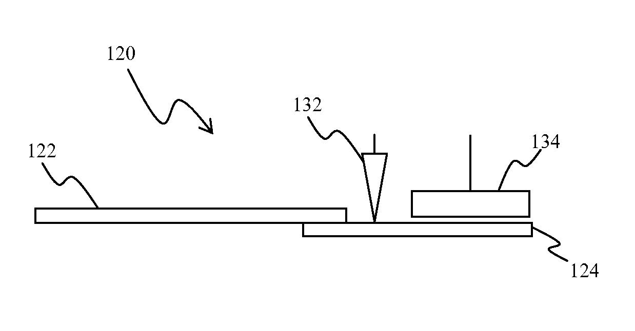
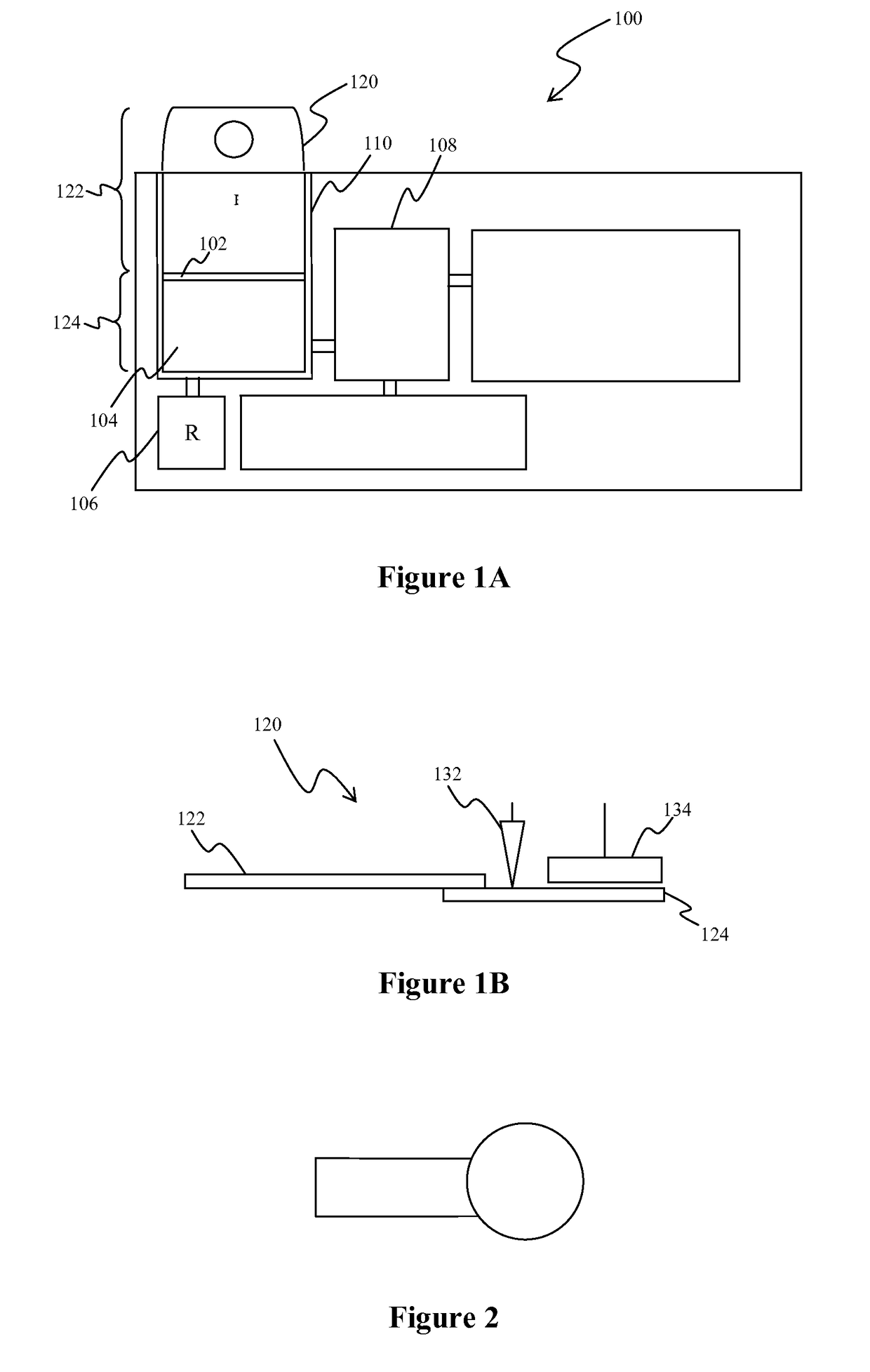
[0042]In a first series of experiments, the sample volumes on the sample pads were determined by weighing the pads before and after sample introduction. The sample pads used were Ahlstrom CYTOSEP 1660 specialty paper and were cut to a specific size of 14 mm by 7 mm and shape as shown in FIG. 2. After sizing the sample pads, plasma was added to a blood separation device to determine the approximate capacity of the sample pad when cut to the above dimensions.

[0043]Table 1 depicts the measurements found when plasma was added to sample pad.

[0044]

TABLE 1Weight 1Weight 2Weight 3Average(mg)(mg)(mg)(mg)Blank Sample Pad12.012.112.0Saturated Sample Pad28.233.430.330.6

[0045]Based on the figures in the table above, the net weight of the sample was 18.6 mg. The precision of the gravimetric measurements for the samples were thus calculated as having a mean of 18.6 mg, standard deviation of 2.62 mg, and a coefficient of variation of 14.1%.

experiment set 2

[0046]In a second series of experiments, a volume was transferred from the sample pad to a TSH immunoassay to determine extraction volume efficiency. This determination was performed by using: (1) sample volumes of 10 μL, 20 μL, and 30 μL directly pipetted into FastPack TSH assay pouches wherein the assay was conducted; and (2) pads containing sample that were sealed into the sample chambers of multiple FastPack TSH pouches wherein the assay was conducted. Six replicates using three different analyzers were used. Moreover, 20 μL of samples were pipetted onto each of 9 sample pads and sealed into the sample chambers of individual FastPack pouches. Based on the above, a TSH standard curve was developed using the sample volumes of 17.5 μL and 20 μL. The extracted volumes were then calculated based upon the chemiluminescent signal generated (RLUs or Relative Light Units). Thus, the average volume of sample extracted was determined to be 17.6 μL and the extracted volume efficiency based ...


PUM

PropertyMeasurementUnit
widthaaaaaaaaaa
widthaaaaaaaaaa
saturation volumeaaaaaaaaaa

Abstract

Devices and methods are presented in which a plasma separation device with a first and second portion separates a blood containing fluid. Most preferably, the first portion produces a cell fraction and a plasma fraction, and the second portion captures the plasma fraction. A first actuator then fluidly isolates a portion of the plasma fraction within the second portion, and a second actuator moves the isolated portion of the plasma fraction from the second portion.

Description

FIELD OF THE INVENTION[0001]The field of the invention is analytical devices, and especially as it relates to devices that include a whole blood separation unit.BACKGROUND OF THE INVENTION[0002]In vitro diagnostic tests to identify and treat diseases have become common tools in hospitals, homes and physician's offices. Biological fluids such as blood, urine or cerebrospinal fluids, which may at times contain blood, are the most frequently employed biological samples for such tests. Of these, blood is the most commonly used. However, for most testing applications, blood must be separated into plasma or serum prior to testing.[0003]Blood separation technologies can generally be grouped within three categories—centrifugation, filtration, and solid-phase separation.[0004]Centrifugation is generally used to separate cellular components from serum or plasma because (1) centrifugation can separate cellular components from serum or plasma at an efficiency of greater than 95%; (2) centrifuge...

Claims


Application Information

Patent Timeline
Patent Type & Authority Patents(United States)
IPC IPC(8): G01N21/75G01N33/49G01N1/40B01D69/02
CPCG01N33/491G01N1/4005B01D69/02B01D2319/06B01D2325/36B01D2325/38
Inventor POIRIER, MICHAELMIDDLETON, JOHN STEPHENPOIRIER, SUZANNE MARIE
Owner QUALIGEN INC
Learn More