System for the internal qualitative and quantitative validation of marker indices
a technology for identifying markers and indicators, applied in the field of qualitative and quantitative validation tools for identifying markers, can solve the problems of difficult commercialization of this procedure, high cost and time consumption of validation methods, etc., and achieve the effect of reliable results
- Summary
- Abstract
- Description
- Claims
- Application Information
AI Technical Summary
Benefits of technology
Problems solved by technology
Method used
Image
Examples
example 1
[0064]MIB-1 immunostaining of pseudo-tissues whose cells consist of 100% of IM9 or Ag8 cell line cells.
[0065]1-3 μm thick sections were stained with the following reagents of the company Monotec, Hamburg, according to the following formula.
Reagents of the MIB-1 Detection Kit
Staining Instructions
De-Paraffination and Rehydration
[0066]Before immunostaining, the paraffin sections applied to the slide must be de-paraffinated in order to remove the embedding medium. Subsequently, the sections must be rehydrated. Insufficient de-parafination and insufficient rehydration are to be avoided by all means, because this can lead to an increased non-specific staining and decreased specific staining.
[0067]
VialAmountDescription11 × 10 mlPeroxidase blocking reagent21 × 5 mlMouse monoclonal antibody MIB-1: ready-to-useantibody purified from culturesupernatant inPBS (0.01 M sodium phosphate, 0.25 MNaCl, pH 7.4-7.6) with stabilizingprotein and sodium azide)Immunogen: bacterially expressed portionsof ex...
example 2
[0104]Immunostaining of pseudo-tissues with defined amounts of AG8 and IM9 cells.
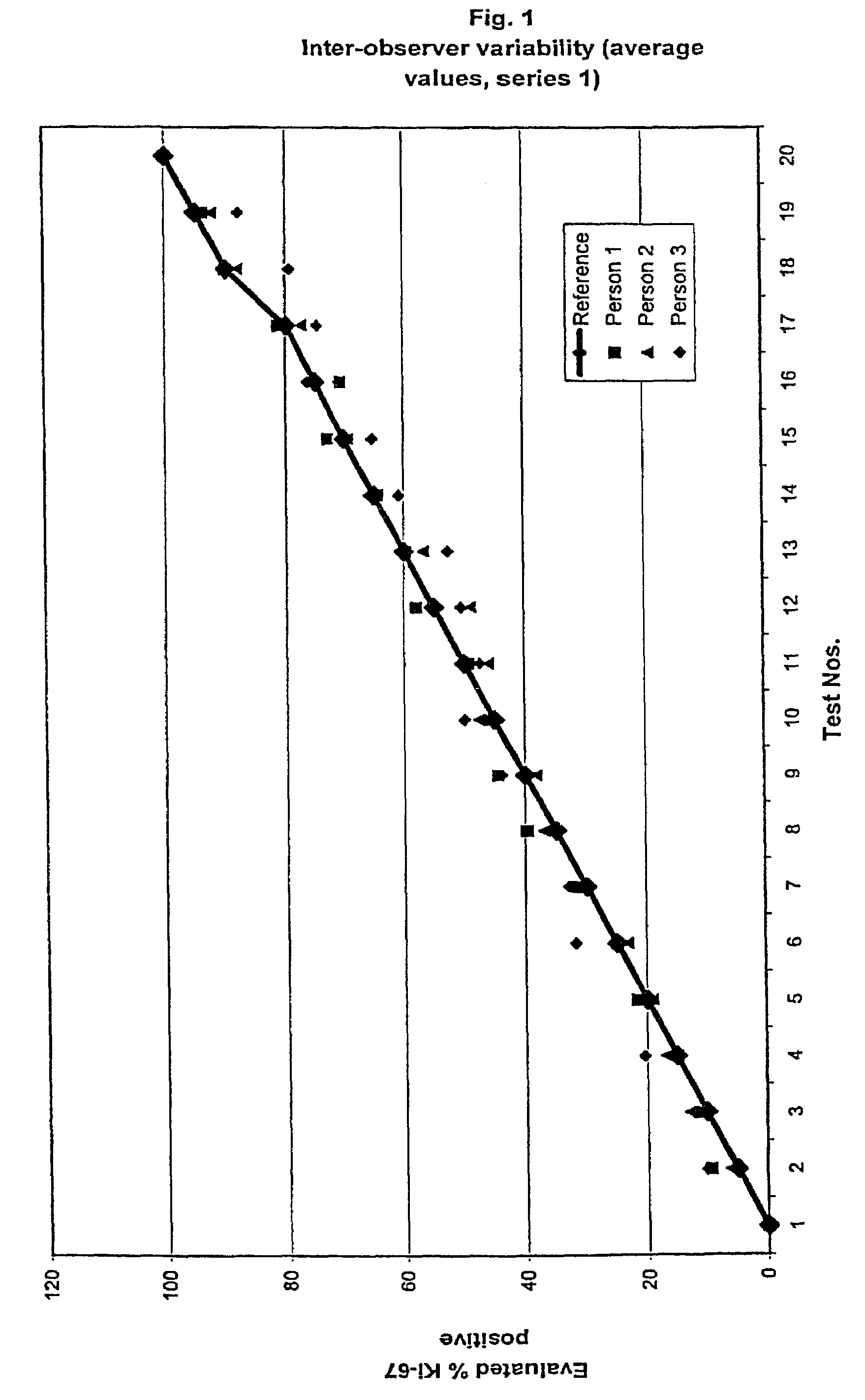
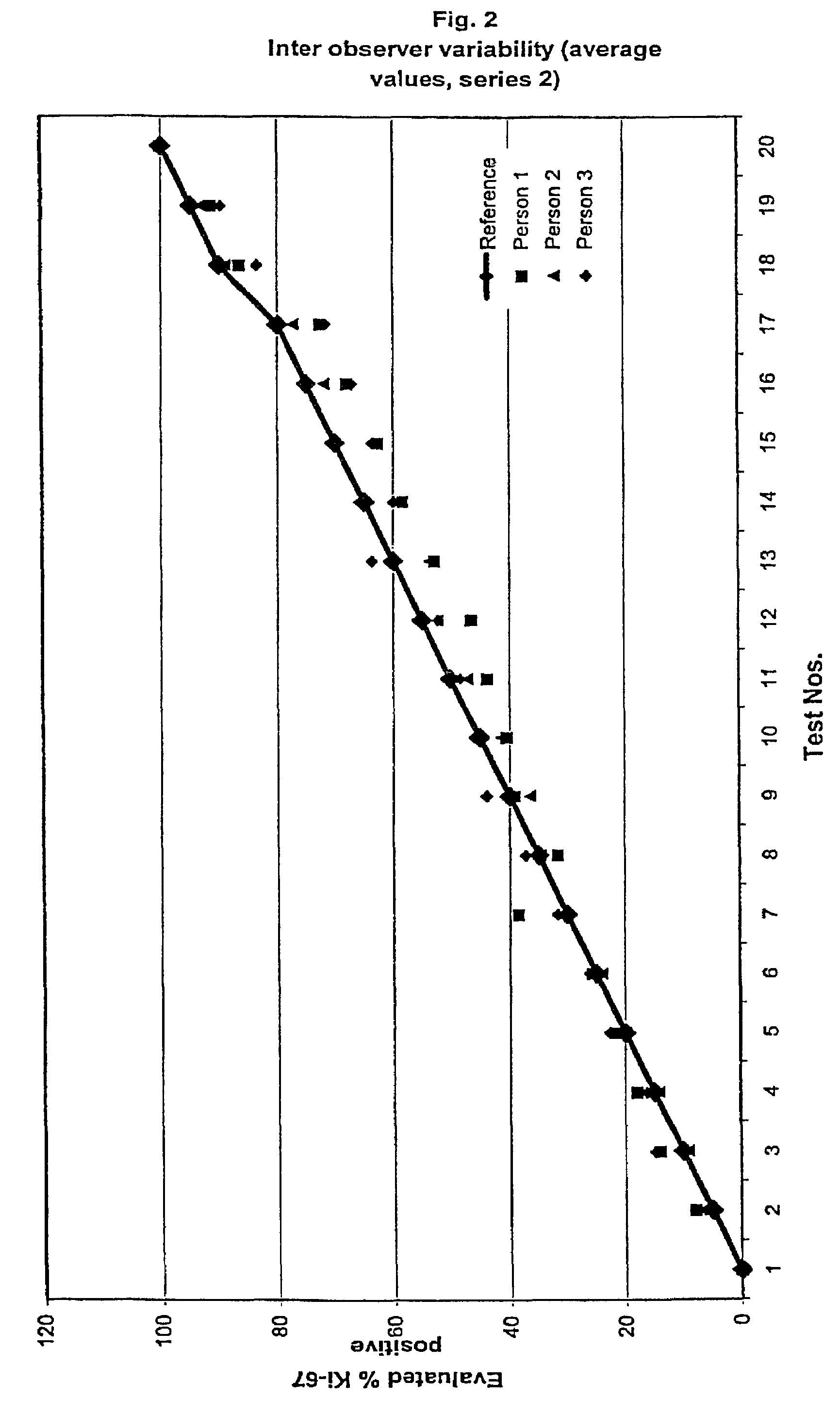
[0105]As depicted above, paraffin blocks of pseudo-tissues were produced with defined amounts of AG8 and / or IM9 cells, respectively, (0, 5, 10, 15, 20, 25, 30, 35, 40, 45, 50, 55, 60, 65, 70, 75, 80, 85, 90, 95 and 100%).
[0106]Paraffin sections were then immunostained as depicted in example 1.
[0107]All stainings with the negative control reagent were negative for all blocks as expected. 0% positive cells. Intra- as well as inter-assay deviation as well as intra- and inter-observer deviations for the negative control reagent were equal to 0.
[0108]Two series were performed.
[0109]The results for the MIB-1 The intra-observer stainings for the two series are summarized in table 1a and 1b and shown in FIGS. 1 and 2, respectively. As it can be observed all raised values are very close to the reference and thereby within the confidence interval.
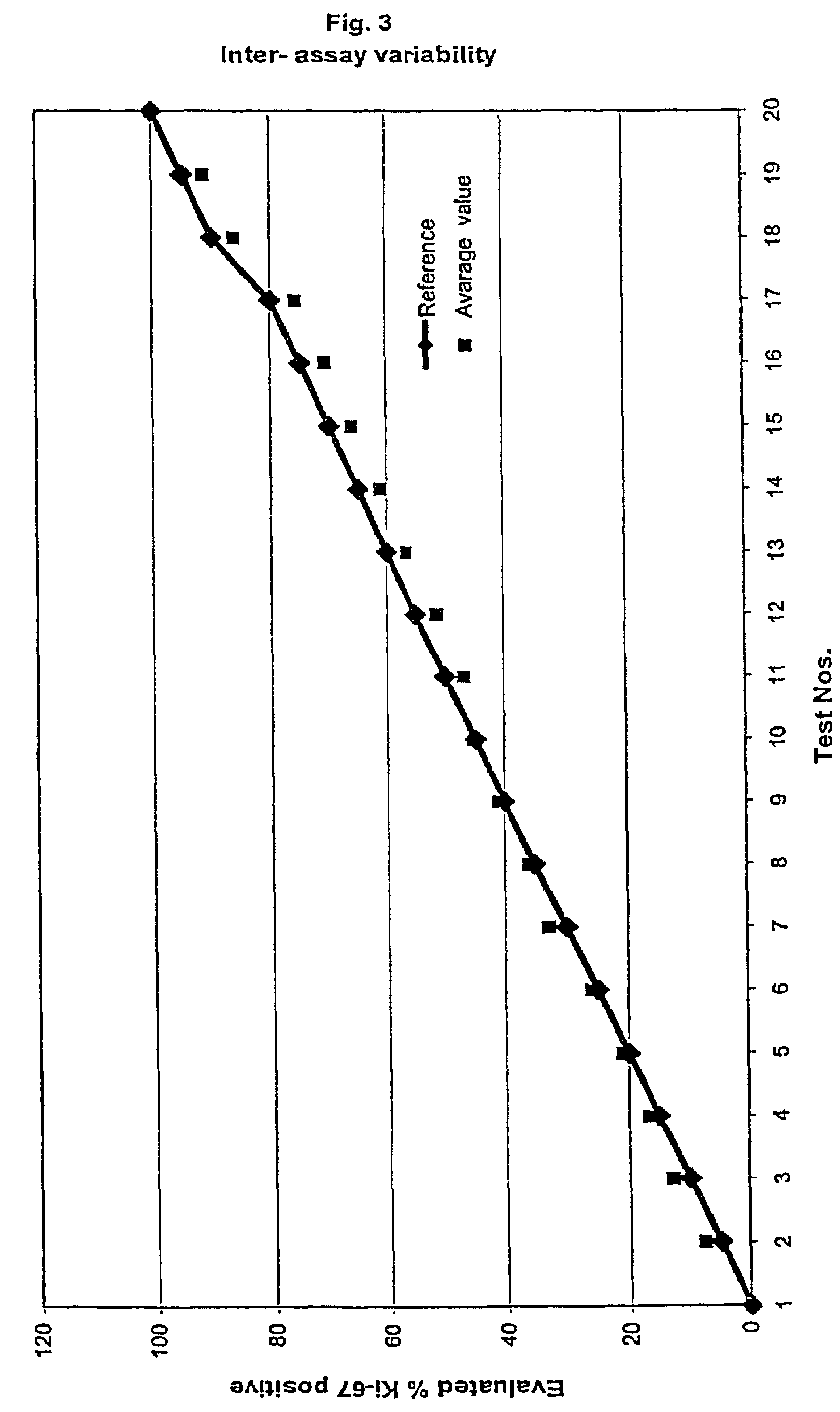
[0110]In table two the average value of the intra-observer test is ...
example 3
Immunostaining of Pseudo-Tissues
[0114]Pseudo-tissues each with a 50% amount of the cell lines Raji (human) and Ag8 (murine) or Jurkat and Ag8 were also produced. The results of MIB-1 staining of sections of these pseudo-tissues were in the same range as portrayed in example 2 for the 50% pseudo-tissue of IM9 and Ag8. Thus, the pseudo-tissue can also be reliably prepared with other cell line cell combinations.
PUM
| Property | Measurement | Unit |
|---|---|---|
| thickness | aaaaa | aaaaa |
| thickness | aaaaa | aaaaa |
| thickness | aaaaa | aaaaa |
Abstract
Description
Claims
Application Information
 - R&D
- Intellectual Property
- Life Sciences
- Materials
- Tech Scout
- Unparalleled Data Quality
- Higher Quality Content
- 60% Fewer Hallucinations
Browse by: Latest US Patents, China's latest patents, Technical Efficacy Thesaurus, Application Domain, Technology Topic, Popular Technical Reports.
© 2025 PatSnap. All rights reserved.Legal|Privacy policy|Modern Slavery Act Transparency Statement|Sitemap|About US| Contact US: help@patsnap.com


