Culture medium for epithelial stem cells and organoids comprising the stem cells

a technology of epithelial stem cells and culture medium, which is applied in the field of culture medium, can solve the problems that no culture system is known that preserves the basic physiology of crypt-villus, and achieve the effect of profound effect on the differentiation of specific cells

Active Publication Date: 2017-09-05
KONINK NEDERLANDSE AKADE VAN WETENSCHAPPEN
View PDF108 Cites 11 Cited by
  • Summary
  • Abstract
  • Description
  • Claims
  • Application Information

AI Technical Summary

Benefits of technology

[0011]The epithelium lining the pyloric region of the small and large bowel encompasses luminal protrusions, villi, and invaginations, crypts. Each cell along the crypt-villus axis is polarized, whereby cells on the top of the intestinal villi, or in the upper positions of colonic crypts, are the most differentiated and are continuously lost into the lumen. Continuous proliferation of stem cells residing in the basis of the crypts, and massive proliferation of progenitor cells residing in the middle of the crypts, ensures proper replacement of the shed cells.
[0325]It will be apparent to a person skilled in the art that the sequential purification steps are not necessarily required to involve the same physical method of separation. Therefore, it will be clear that, for example, the cells may be purified through a FACS step using an anti-Lgr5 antibody, followed by an immuno-affinity purification step using a SSEA-1 affinity column. In certain embodiments, the cells may be cultured after isolation for at least about 15, at least about 20 days, at least about 25 days, or at least about 30 days. In certain aspects, the cells are expanded in culture longer to improve the homogeneity of the cell phenotype in the cell population.

Problems solved by technology

Furthermore, no culture system is known that preserves the basic crypt-villus physiology of crypts that have been isolated from colon or intestine, or that preserves the basic physiology of isolated pancreatic fragments or gastric tissue fragments.

Method used


Image

  • Culture medium for epithelial stem cells and organoids comprising the stem cells
  • Culture medium for epithelial stem cells and organoids comprising the stem cells
  • Culture medium for epithelial stem cells and organoids comprising the stem cells

Examples


example 1

of Small Intestine Crypts and Villi in Vitro

Materials and Methods

[0401]Mice: Outbred mice of 6-12 weeks of age were used. Generation and genotyping of the Lgr5-EGFP-Ires-CreERT2 allele1 has been previously described1. Rosa26-lacZ or YFP Cre reporter mice were obtained from Jackson Labs.

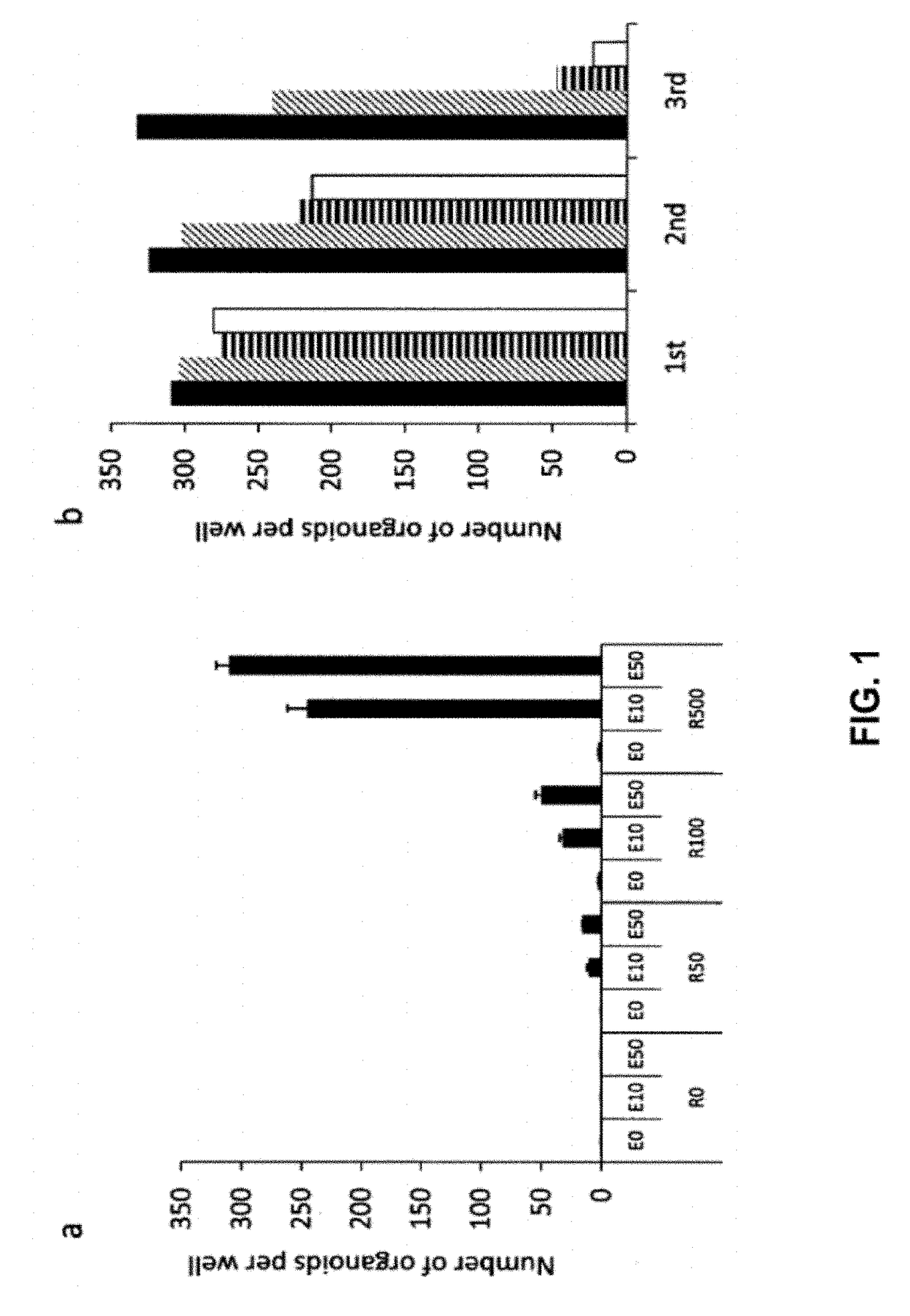
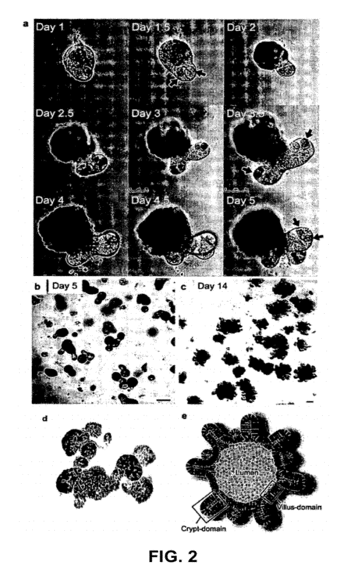
[0402]Crypt isolation, cell dissociation and culture: Crypts were released from murine small intestine by incubation in 2 mM EDTA / PBS for 30 min at 4° C. Isolated crypts were counted and pelleted. 500 crypts were mixed with 50 μl Matrigel (BD Bioscience) and plated in 24-well plates. After polymerization of Matrigel, 500 μl of crypt culture medium (Advanced DMEM / F12 with growth factors (10-50 ng / ml EGF (Peprotech), 500 ng / ml R-spondin 111 and 100 ng / ml Noggin (Peprotech)) was added. For sorting experiments, isolated crypts were incubated in culture medium for 45 min at 37° C., following by resuspension with a glass pipette. Dissociated cells were passed through 20-μm cell strainer. GFPhi, GFPlow or GF...

example 2

of Colon Crypts and Villi in Vitro

Material and Methods

Wnt3a Conditioned Medium

[0420]A Wnt3a ligand expressing cell line and the same cell line, without the Wnt3a ligand (control medium) are cultured for a period of 3-4 weeks. The cells will produce Wnt3a as soon as they stop grown at confluency. The medium will be harvested and tested in the TOPflash assay, a luciferase assay using a TCF responsive elements-luc construct (TOP) and the same construct, but with mutations in the TCF responsive elements (FOP). The ratio between TOP / FOP should be more than 20 for the medium to be used in cultures. The medium is diluted 25-50% when used in the cultures to regenerate tissue.

[0421]Freshly isolated colon was opened and washed with PBS or DMEM, and cut into small pieces. The fragments were incubated with 2 mM EDTA / PBS for 1 hour at 4° C. under gentle rocking. Following removal of EDTA solution, the tissue fragments were vigorously suspended in 10 ml of cold PBS with a 10 ml pipette. The first...

example 3

of Adenomas in Vitro

Materials and Methods

[0424](See example 1)

Results

[0425]Adenomas have been historically difficult to culture in vitro. Since the above described conditions were used to successfully culture healthy crypts derived from small intestine as well as colon, it was determined whether similar conditions could sustain adenomas in vitro. After isolation of adenoma from APC− / − mice using 2.5 mM EDTA, single adenomas were cultured under similar conditions as described above. Importantly, these conditions were adequate to maintain growth of the adenomas in vitro, however, R-spondin had become redundant. This can be easily explained by the fact that it no longer is necessary to induce the Wnt signaling pathway, since the absence of APC in these cells will automatically result in nuclear β-Catenin. This makes R-spondin, a Wnt agonist, redundant in culturing adenomas in vitro. FIG. 16a, and in larger magnification in FIG. 16b, show that, in contrast to normal crypt organoids, in ...


PUM


Abstract

The invention relates to a method for culturing epithelial stem cells, isolated tissue fragments comprising the epithelial stem cells, or adenoma cells, and culturing the cells or fragments in the presence of a Bone Morphogenetic Protein (BMP) inhibitor, a mitogenic growth factor, and a Wnt agonist when culturing epithelial stem cells and isolated tissue fragments. The invention further relates to a cell culture medium comprising a BMP inhibitor, a mitogenic growth factor, and a Wnt agonist, to the use of the culture medium, and to crypt-villus organoids, gastric organoids, pancreatic organoids, liver organoids, colon organoids, Barrett's Esophagus organoids, adenocarcinoma organoids and colon carcinoma organoids that are formed in the culture medium.

Description

CROSS-REFERENCE TO RELATED APPLICATIONS[0001]This application is a continuation-in-part of co-pending application U.S. Ser. No. 13 / 147,163, filed Sep. 14, 2011, which is the national stage under 35 U.S.C. §371 of International Application Number PCT / NL2010 / 000017, filed Feb. 3, 2010, designating the United States of America, and published, in English, on Aug. 12, 2010 as WO 2010 / 090513, which claims priority under 35 U.S.C. §119(e) to U.S. provisional patent application Ser. No. 61 / 149,622, filed Feb. 3, 2009, and further claims priority under Article 8 of the PCT to European Patent Office applications EP 09151970.2, filed Feb. 3, 2009, and EP 09171831.2, filed Sep. 30, 2009. This application also claims the benefit under 35 U.S.C. §119(e) of U.S. Ser. No. 61 / 368,736, filed on Jul. 29, 2010, U.S. Ser. No. 61 / 520,569, filed on Jun. 10, 2011, and U.S. Ser. No. 61 / 571,663, filed Jun. 30, 2011. This application also claims priority under 35 U.S.C. §119 to European Patent Office applicat...

Claims


Application Information

Patent Timeline
Patent Type & Authority Patents(United States)
IPC IPC(8): C12Q1/02C12N5/071C12N5/02
CPCC12N5/0671C12N5/068C12N5/0677C12N5/0679C12N2500/32C12N2500/38C12N2501/11C12N2501/117C12N2501/119C12N2501/12C12N2501/13C12N2501/15C12N2501/155C12N2501/335C12N2501/345C12N2501/415C12N2501/998C12N2533/90
Inventor SATO, TOSHIROCLEVERS, JOHANNES C.HUCH ORTEGA, MERITXELL
Owner KONINK NEDERLANDSE AKADE VAN WETENSCHAPPEN
Why Eureka
Try Eureka