Method and magnetic resonance apparatus for image acquisition control with administration of contrast agent

a technology of contrast agent and magnetic resonance apparatus, which is applied in the field of magnetic resonance apparatus for image acquisition control with can solve the problems of examination having to be shifted, image data set overall poor quality, and too early administration of contrast agent, etc., and achieve the effect of increasing image quality

Active Publication Date: 2017-09-26
SIEMENS HEALTHCARE GMBH
View PDF4 Cites 0 Cited by
  • Summary
  • Abstract
  • Description
  • Claims
  • Application Information

AI Technical Summary

Benefits of technology

[0012]An object of the present invention is to provide a method to control the image acquisition operation in a measurement with contrast agent that enables an increase in the image quality while avoiding the disadvantages of the cited methods of the prior art.
[0021]The basis of the present invention is thus to ultimately combine the test bolus method and automatic detection of the main bolus (bolus tracking) in order to achieve the advantages of both measurement methods. For this purpose, the wait period between the injection of the main bolus and the start of the acquisition protocol (which is present in the test bolus method) is filled with measurements (data acquisitions) of second magnetic resonance images that have a high temporal resolution. As in a procedure with automatic detection of the main bolus, in these second magnetic resonance images it can ultimately be checked whether an early administration of the contrast agent has taken place. Because the wait period would be present anyway, there are no disadvantages to filling these with the measurements of the second magnetic resonance images. The primary advantage of the procedure according to the invention results from this: that the robustness of the test bolus method is increased because a start of the measurement that is markedly too late is avoided without disadvantages being creating in the procedure. In addition, costs are reduced because measurements must be repeated in fewer patients. A high image quality is thereby maintained that can very exactly indicate the peak time at which the highest concentration of contrast agent is present in the acquisition area of the image data set, such that the start point in time of the acquisition protocol can be selected correctly in order to acquire the center of k-space at this time. If a time offset occurs once between the injection of the contrast agent and the beginning of the correspondingly determined wait period, a monitoring is implemented that ensures that the deviation is not too great, and consequently the image quality is increased even given such deviations, in particular because venous contaminations of arterial image data sets can be avoided, even if the peak time and the measurement time of the k-space center do not exactly coincide given a detection of the main bolus in the second magnetic resonance images.
[0023]Overall, an improvement is thus provided both with regard to the purely automatic bolus tracking and with regard to a pure test bolus method. In most cases according to conventional procedures, the precise test bolus measurement is based on the start of the acquisition protocol in which too early an arrival of the contrast agent in the acquisition area of the image data set occurs (be it due to an operator error or due to a physiological variation in the patient), thus resulting in a start of the acquisition protocol that is also too early. This is avoided in the inventive procedure. The dependency of the test bolus measurement on the physiological parameters of the patient is thus likewise reduced.
[0024]In a further embodiment of the present invention, the end of the monitoring interval is selected so that the time period from the end of the monitoring interval until the end of the wait period is shorter than or identical to the acquisition time for a second magnetic resonance image. In this way it can be ensured that the acquisition of the second magnetic resonance images does not affect the coincidence of the k-space center and the peak of the main bolus, such that in each case the acquisition protocol for acquisition of the image data set can be started at the correct point in time after the wait period.
[0030]In a further embodiment of the present invention, the second acquisition area is determined under consideration of image data of the first magnetic resonance images. This means that, in the first magnetic resonance images, the test bolus is observed over a longer time period and / or distance, such that a suitable position and / or orientation (thus attitude) of the second acquisition area can be determined in the target area in order to detect the arrival of the contrast agent as early as possible and—in the event that this occurs before the expiration of the wait period—to be able to start the acquisition protocol for the image data set correspondingly early. A synergy effect is thus used that is achieved via the use of both methods, thus the test bolus measurement and automatic bolus tracking.
[0031]A magnetic resonance sequence can be used for the acquisition of the second magnetic resonance images (which are in particular to be acquired as two-dimensional slices), which magnetic resonance sequence has an acquisition duration per second magnetic resonance image of less than two seconds, in particular less than one second, and / or the length of the monitoring interval can be selected so that at least five second magnetic resonance images (in particular at least 10 second magnetic resonance images) can be acquired in said monitoring interval. For the acquisition of the second magnetic resonance images, a magnetic resonance sequence with high temporal resolution is thus used that enables an optimally fast determination of an arrival of the main bolus in the target area. Suitable sequences are widely known in the prior art, wherein it is noted that ultimately the same basic sequence can be used for the acquisition of the first magnetic resonance images, the second magnetic resonance images and the image data set, wherein such a basic sequence is typically optimized for contrast agent measurements and can be parameterized in order to achieve a higher temporal resolution (for the first and second magnetic resonance images) or a higher spatial resolution (measurement of the image data set).

Problems solved by technology

However, a number of problems occur in a test bolus measurement.
It is possible that, due to a human error, the contrast agent is administered too early, for example, by a start signal for the wait period being provided with a time offset toward the injection of the contrast agent of the main bolus.
This problem exists because the synchronization between the injection of the contrast agent and the start of the wait period must occur manually on the part of the user because, although contrast agent injectors that can automatically trigger the start of a wait period are known in computed tomography systems, an automatic injection controller for magnetic resonance systems is not yet known because it is extremely complicated to produce magnetic resonance-compatible devices.
If the contrast agent is administered too early—meaning before the beginning of the wait period—this has the result that the acquisition protocol (and thus the measurement of the image data set) begins too late, such that the image data set overall has a poorer quality, in particular because venous portions can already be present in the arterial imaging and the like.
In the worst case, the examination must be shifted to a further day because the amount of contrast agent that can be administered to a patient within a given time duration is limited.
An additional problem is the possibility of a variation of the physiological situation of the patient.
For example, if the patient is excited, adrenalin can be produced, which increases the circulation speed so that it may occur, due to such physiological causes, that the contrast agent arrives before the wait period has ended, such that a non-optimal image data set is acquired.
A first intrinsic problem of this variant is the selection of the second acquisition area, which does not correspond to the acquisition area of the image data set (and naturally also does not correspond to the first acquisition area in a test bolus measurement).
The estimation of a suitable second acquisition area is extremely difficult, and as a consequence a further problem of automatic bolus detection results from the poor time resolution of this method because, for example, data of new magnetic resonance images are present only every second or every two seconds.
This poor time resolution, and the fact that the time that the blood (and thus the contrast agent bolus) requires in order to arrive from the second acquisition area to the acquisition area of the image data set is very short, have the result that compromises are often necessary in the quality of the image data sets.

Method used


Image

  • Method and magnetic resonance apparatus for image acquisition control with administration of contrast agent
  • Method and magnetic resonance apparatus for image acquisition control with administration of contrast agent
  • Method and magnetic resonance apparatus for image acquisition control with administration of contrast agent

Examples


Embodiment Construction

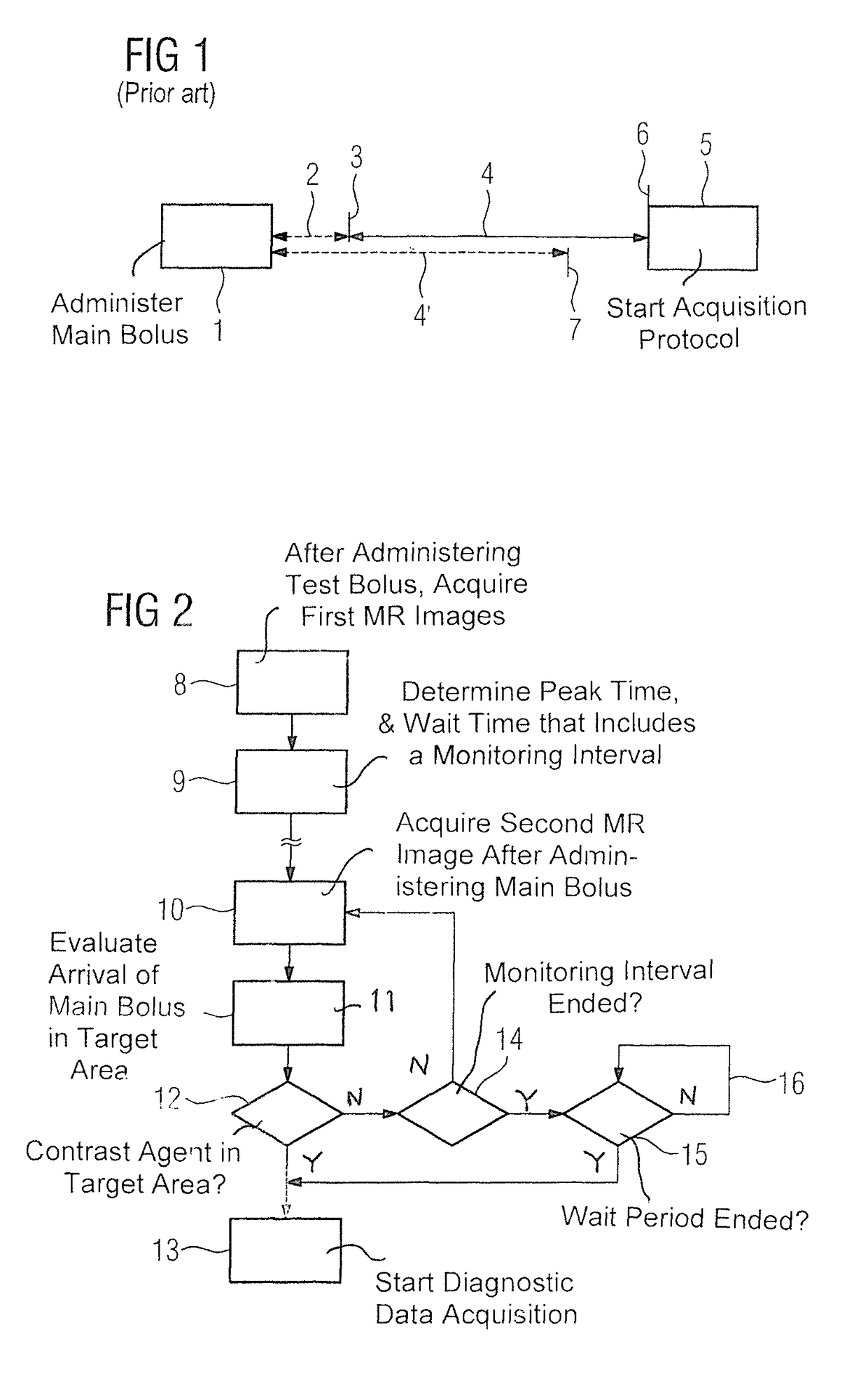
[0038]FIG. 1 explains a problem underlying the present invention in an example of a test bolus measurement according to the prior art. This involves the acquisition of an image data set with a magnetic resonance device, wherein it is thereupon sought that the k-space center coincides in agreement with a peak time in which the concentration of the contrast agent in the acquisition area of the image data set is highest. A smaller amount of contrast agent—what is known as the test bolus—is consequently administered in the test bolus method (as it is known from the prior art) than in the main bolus. First magnetic resonance images from which the peak time can be determined are acquired using a magnetic resonance sequence of high temporal resolution. With these images, a wait period is calculated for the main bolus with the measurement of the k-space center in that the time from the start of the acquisition protocol for the image data set up to the measurement of the k-space center is su...


PUM


Abstract

In a method and magnetic resonance apparatus to acquire diagnostic image data of a contrast agent-filled target area of a patient, a peak time of the test bolus in the target area is automatically determined, from which a wait period is then determined for administering the main bolus. After the main bolus has been administered to the patient, magnetic resonance images of the target area are acquired, and each is analyzed immediately after acquisition thereof to determine whether that image shows arrival of the contrast agent. If and when one of these images shows such arrival, an acquisition protocol is immediately started in order to acquire the diagnostic image data set. If none of these images shows arrival of the contrast agent, the protocol to acquire diagnostic image data is started after the wait period.

Description

BACKGROUND OF THE INVENTION[0001]Field of the Invention[0002]The present invention concerns a method to control the acquisition of a diagnostic image data set of at least one part of a contrast agent-filled target area of a patient with a magnetic resonance device according to an acquisition protocol, as well as a magnetic resonance apparatus designed to perform such a method.[0003]Description of the Prior Art[0004]Contrast agent is often used within the scope of magnetic resonance imaging, in particular when image exposures should be produced within the context of dynamic processes, and for perfusion data acquisition in the vascular system of a patient. One important sign of quality of the image data sets that are acquired in this manner is that a good contrast agent filling is present. This means that the acquisition of the image data set must be implemented so that the acquisition region is optimally filled with contrast agent during the entire acquisition time. Magnetic resonanc...

Claims


Application Information

Patent Timeline
Patent Type & Authority Patents(United States)
IPC IPC(8): G01R33/56A61B5/055A61B5/026
CPCG01R33/5601A61B5/0263A61B5/055
Inventor RICK, MANUELASCHMITT, PETERDE OLIVEIRA, ANDRE
Owner SIEMENS HEALTHCARE GMBH
Why Eureka
Try Eureka