

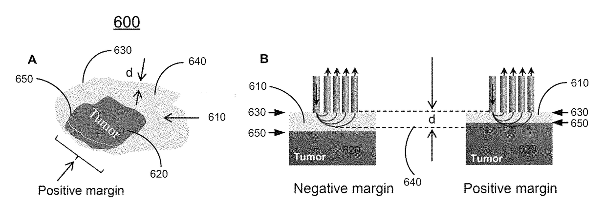
Methods and systems for three-dimensional real-time intraoperative surgical margin evaluation of tumor tissues
a tumor tissue and intraoperative technology, applied in the field of surgical procedures, can solve the problems of increasing patient morbidity and healthcare costs, reducing patient survival rates, and resected specimens being strongly correlated with the risk of local tumor recurren
- Summary
- Abstract
- Description
- Claims
- Application Information
AI Technical Summary
Benefits of technology
Problems solved by technology
Method used
Image

Examples
example one
[0165]In this example, in vitro study is performed. Preliminary Raman and fluorescence spectra have been acquired to (1) demonstrate the ability of Raman and fluorescence spectroscopy in differentiating STS and normal tissues and (2) confirm the feasibility of measuring Raman and fluorescence signal of tumor beds in the operating room.
[0166]Under VU IRB approval (#120813), tumor and control samples from 20 patients have been acquired from the VU Cooperative Human Tissue Network. Raman spectra of the samples were collected using a portable fiber optic system, yielding 44 spectra from tumors and 33 spectra from control muscles.
[0167]FIG. 10A shows schematically average Raman spectra of control muscle and STS samples according to certain embodiments of the present invention. Multivariate statistical analysis was used to classify spectra into tumor and control groups with 100% sensitivity and 100% specificity, as shown in Table 2.
[0168]
TABLE 2Classification matrix for in vitro specimens...
example two
[0172]In this example, pilot clinical study is performed. The initial work demonstrated the feasibility of using Raman and fluorescence spectroscopy to differentiate STS and muscle on intact specimens in a laboratory setting. Studies are currently underway using the same approach in a clinical setting, and results are equally promising. Raman spectra were recorded for 3-10 seconds from multiple sites in the tumor bed of 19 patients undergoing STS excision at the VU Medical Center.
[0173]FIG. 13 shows schematically Raman spectra of in vivo tissues according to certain embodiments of the present invention. As shown in FIG. 13, spectrum from 1 to 8 is the Raman spectrum of healthy tissues, as data 1 represents synovium; data 2 represents tendon; data 3 represents skin; data 4 represents nerve; data 5 represents bone marrow; data 6 represents fat; data 7 represents bone; and data 8 represents muscle. The Raman spectra of STS tumors from selected 4 patients are marked as p1-4 and are hist...
PUM
| Property | Measurement | Unit |
|---|---|---|
| depth resolution | aaaaa | aaaaa |
| optical data | aaaaa | aaaaa |
| morphological surface | aaaaa | aaaaa |
Abstract
Description
Claims
Application Information