Method for multi-layer spiral CT three-dimensional rebuilding measuring humeral head eccentricity
A technology of three-dimensional reconstruction and humeral head, applied in echo tomography, computerized tomography scanner, etc., can solve problems such as difficult observation of humeral shaft, achieve objective data and improve accuracy
- Summary
- Abstract
- Description
- Claims
- Application Information
AI Technical Summary
Problems solved by technology
Method used
Image
Examples
Embodiment 1
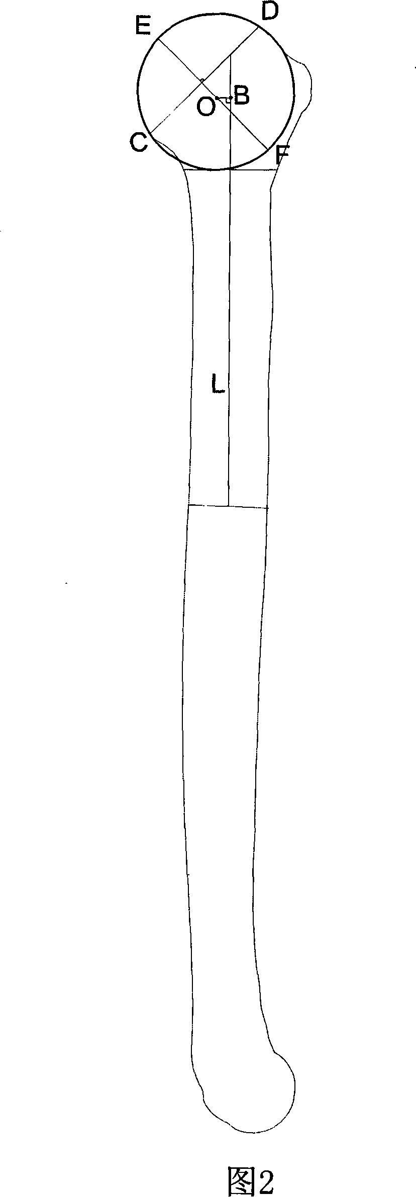
[0026] As shown in Figures 1 and 2, the present invention discloses a method for measuring humeral head eccentricity by multi-slice spiral CT three-dimensional reconstruction, comprising the following steps:
[0027] 1. The method for measuring humeral head eccentricity by multi-slice spiral CT three-dimensional reconstruction comprises the following steps:
[0028] (1) The humerus specimen is placed on the examination bed, and the orthogonal positioning line is located on the front and side mid-axis of the humerus respectively;
[0029] (2) CT scan: Use a multi-slice spiral CT scanner to collect continuous scan data of the humerus specimen. The CT scan parameters are 120KV, effective mAs150, collimator width 0.75mm, acquisition layer thickness 5mm, overlap 2mm, the CT scan range is from the highest point of the humeral head to the end of the humeral trochlear;
[0030] (3) Three-dimensional reconstruction: the humerus is reconstructed by volume reconstruction imaging process...
Embodiment 2
[0034] The invention discloses a method for measuring humeral head eccentricity by multi-slice spiral CT three-dimensional reconstruction, comprising the following steps:
[0035] 1. The method for measuring humeral head eccentricity by multi-slice spiral CT three-dimensional reconstruction comprises the following steps:
[0036] (1) Shoulder joint specimens are placed on the examination table, with the orthogonal positioning lines respectively located on the front and side mid-axis of the humerus;
[0037] (2) CT scan: use multi-slice spiral CT scanner to carry out continuous scan data collection on the shoulder joint, CT scan parameters are 130KV, effective mAs140, collimator width 1.5mm, acquisition layer thickness 5mm, overlap 2mm; its CT scan range is from the acromion to the end of the trochlear of the humerus.
[0038] (3) Three-dimensional reconstruction: the shoulder joint is reconstructed by volume reconstruction imaging, and the scapula needs to be cut and separate...
PUM
 Abstract
Description
Claims
Application Information
 - R&D
- Intellectual Property
- Life Sciences
- Materials
- Tech Scout
- Unparalleled Data Quality
- Higher Quality Content
- 60% Fewer Hallucinations
Browse by: Latest US Patents, China's latest patents, Technical Efficacy Thesaurus, Application Domain, Technology Topic, Popular Technical Reports.
© 2025 PatSnap. All rights reserved.Legal|Privacy policy|Modern Slavery Act Transparency Statement|Sitemap|About US| Contact US: help@patsnap.com


