Breast image processing system and breast image processing method

An image processing and breast technology, applied in the field of breast image processing systems, can solve the problems of transparent parts, lack of image information, difficulty in observing the chest wall, etc., and achieve the effect of preventing image loss

Inactive Publication Date: 2008-03-12
KONICA MINOLTA MEDICAL & GRAPHICS INC
View PDF3 Cites 2 Cited by
  • Summary
  • Abstract
  • Description
  • Claims
  • Application Information

AI Technical Summary

Problems solved by technology

[0007] However, because of the JIS standard, the film size has an allowable error of ±1mm. For example, the relationship between the exposure scanning start position (writing start position) H and the image, as shown in Figure 18, when the film size has a + (positive) error , the film size is larger than the data size of the image data, so, as shown in FIG. 18(a), a transparent portion that is not exposed to the laser appears on the chest wall side.
When observed on the reading box, strong light passes through the transparent part, which makes the observer feel dazzling and makes it difficult to observe the chest wall, so it is not ideal
In addition, if there is a - (negative) error in the film size and the film size is smaller than the data size of the image data, then as shown in FIG. 18( b ), the image information of the chest wall part important for diagnosis is lost, which also becomes a problem.
In addition, when the position limit of the film deviates slightly, transparent parts may also occur, resulting in defects such as loss of image information on the chest wall.

Method used


Image

  • Breast image processing system and breast image processing method
  • Breast image processing system and breast image processing method
  • Breast image processing system and breast image processing method

Examples


Embodiment Construction

[0107] First, the structure of this embodiment will be described.

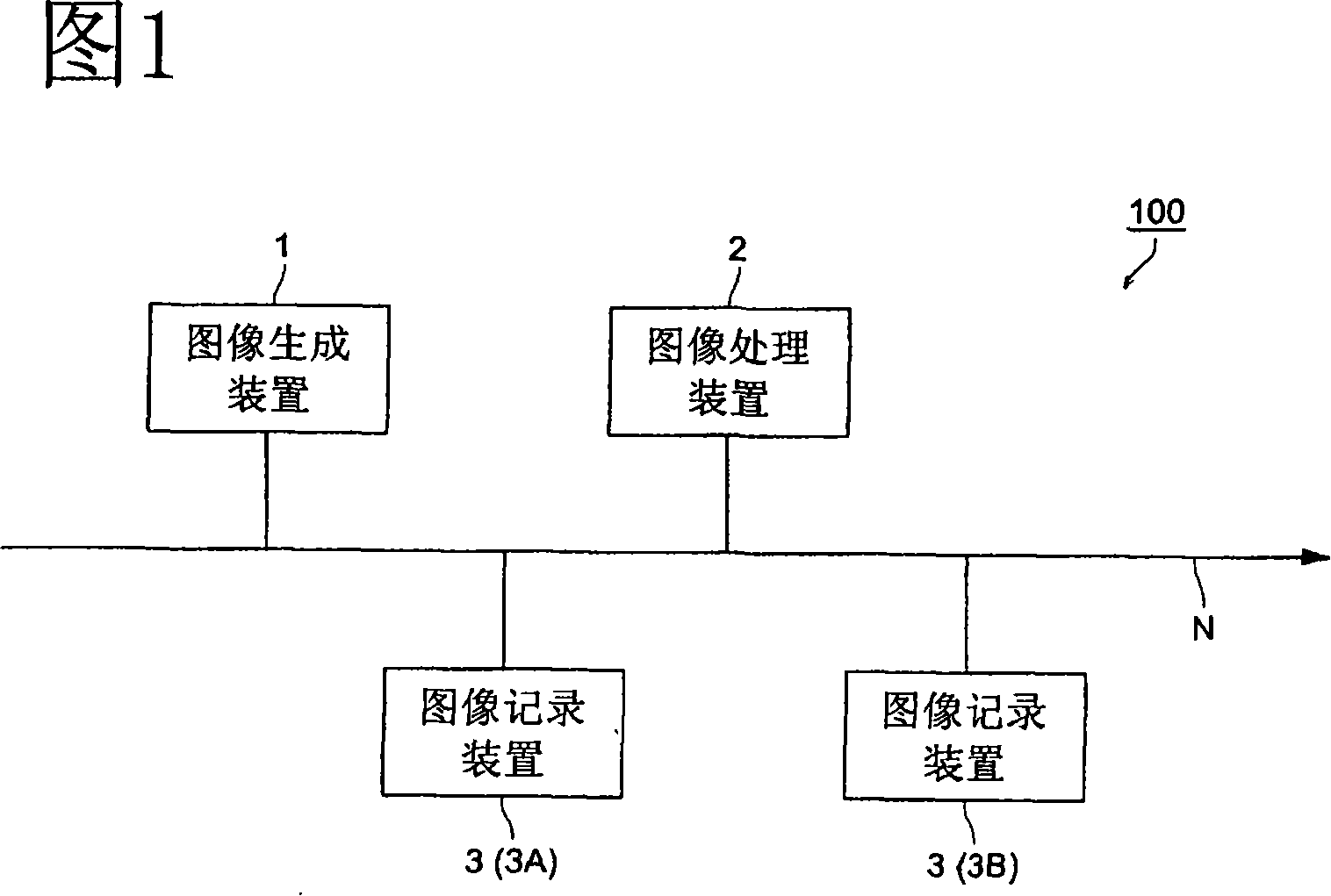
[0108] FIG. 1 is a conceptual schematic diagram of the overall structure of a breast image processing system 10 according to this embodiment. As shown in FIG. 1 , a breast image processing system 100 is an image generating device 1, an image processing device 2, an image recording device 3, etc. connected through a communication network N (hereinafter referred to as network N), and can transmit and receive data mutually.

[0109] In this embodiment, an example of connecting the image generating device 1, the image processing device 2, and the image recording device 3 via a network is described, but the present invention is not limited thereto, and a system structure in which the devices are directly connected by wires may also be used. In addition, the number and installation place of each device are not particularly limited, either.

[0110] The network N can be in various forms such as LAN (Local Area Netwo...


PUM


Abstract

The problems that a skipping area is shown in the chest wall portion of the output image and image information on the chest wall portion lacks when the left and right breast images are recorded on films and outputted to compare and examine the images on the film viewer are solved. An image processing device of a breast image processing system performs formatting so that the chest wall portions of left and right breast images created by an image creating device may be disposed on the write start position H sides of an image recording device.

Description

technical field [0001] The present invention relates to a breast image processing system and a breast image processing method, wherein the left and right breast images are formatted and recorded separately on a recording medium for output. Background technique [0002] In the past, medical images input as digital images by CT (Computed Tomography), MRI (Magnetic Resonance Imaging), CR (Computed Radiography), FPD (Flat Panel Detector), ultrasonic diagnostic equipment, etc. After various image processing including processing, it is displayed on a monitor such as a CRT (Cathode Ray Tube), and the doctor performs reading observation on the monitor. Or after performing various image processing, use an image recording device to record the medical image on a recording medium such as a film, and output it in hard copy, and the doctor will perform the reading and observation on the viewing box. [0003] When a doctor reads a medical image of a breast (mammography), he ordinarily com...

Claims


Application Information

Patent Timeline
Patent Type & Authority Applications(China)
IPC IPC(8): A61B6/00G06T3/00
CPCG06T2207/10116A61B6/502G06T2207/30068G06T7/0028G06T7/33
Inventor 山道洋次赤木英一梅木守
Owner KONICA MINOLTA MEDICAL & GRAPHICS INC
Why Eureka
Try Eureka