Pet/mr scanner with time-of-flight capability

A scanner and imaging system technology, applied in the field of imaging technology, can solve problems such as high noise and low radiation count rate of detectors, and achieve the effect of simplifying the structure

Active Publication Date: 2008-04-16
KONINKLIJKE PHILIPS ELECTRONICS NV
View PDF0 Cites 24 Cited by
  • Summary
  • Abstract
  • Description
  • Claims
  • Application Information

AI Technical Summary

Problems solved by technology

However, smaller detectors have lower radiation count rates and correspondingly higher noise

Method used


Image

  • Pet/mr scanner with time-of-flight capability
  • Pet/mr scanner with time-of-flight capability
  • Pet/mr scanner with time-of-flight capability

Examples


Embodiment Construction

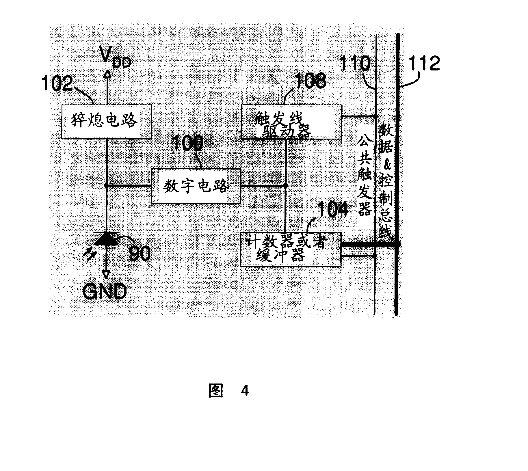
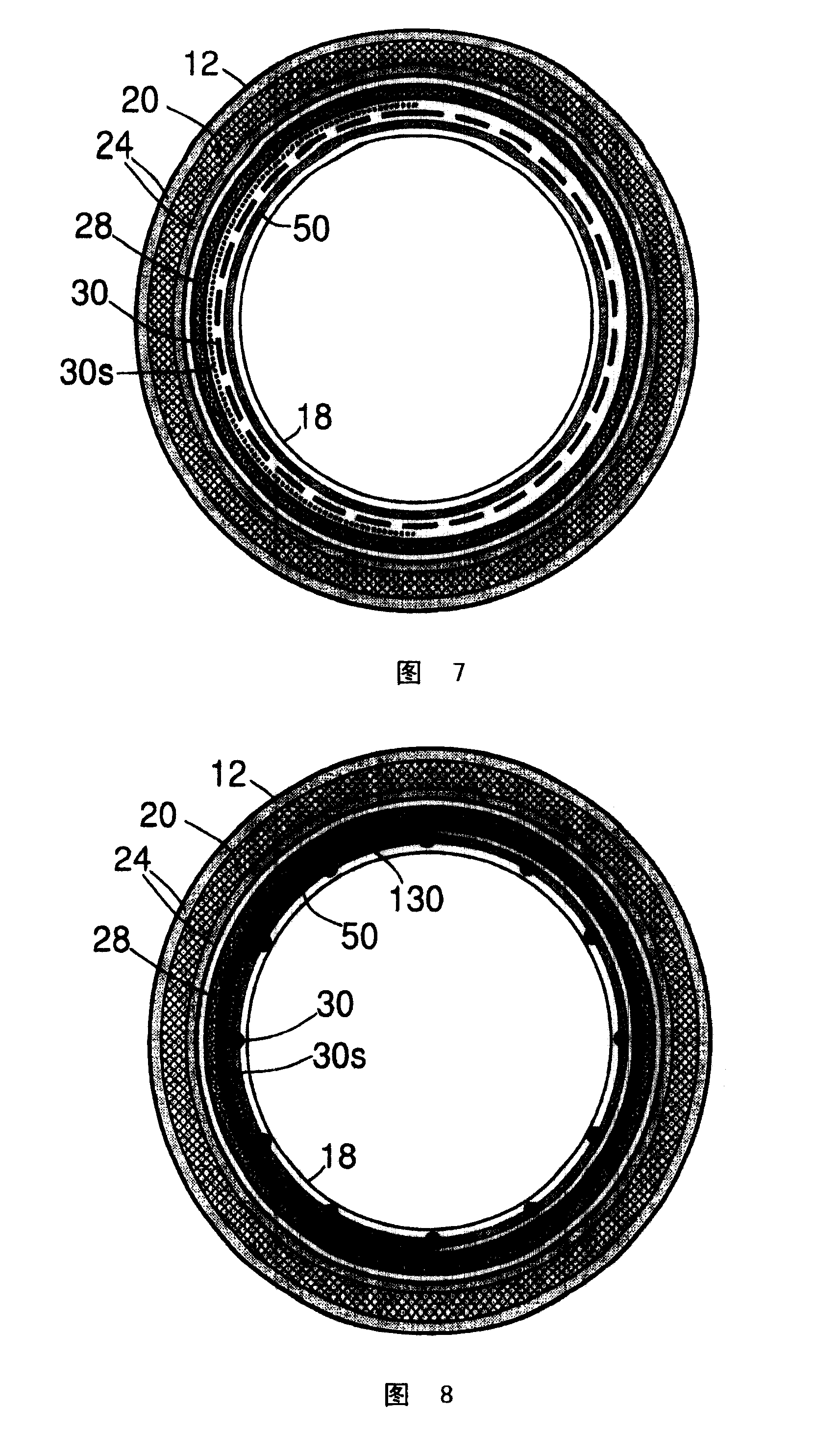
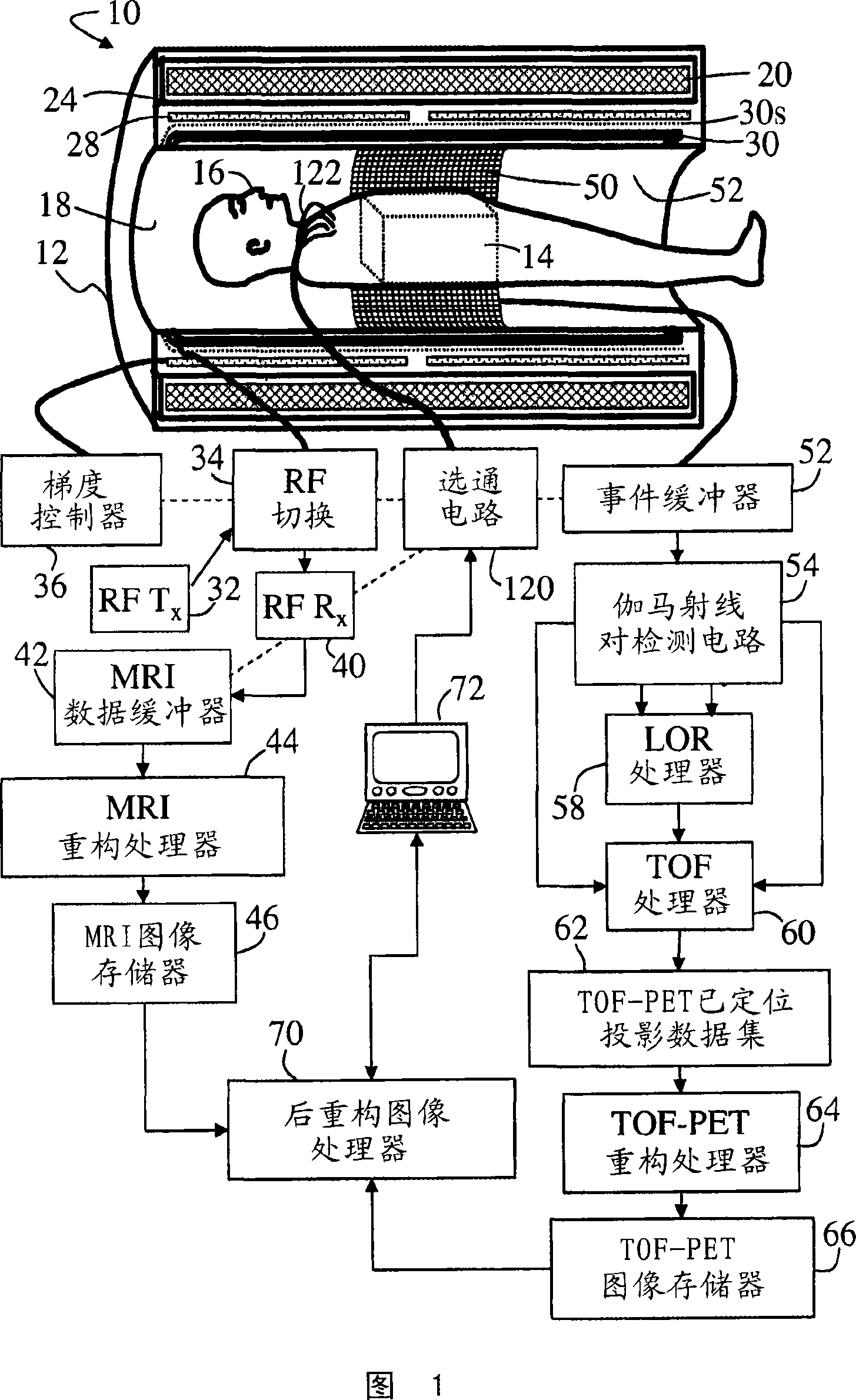
[0023] Referring to FIG. 1 , a combined positron emission tomography / magnetic resonance imaging (PET / MRI) scanner 10 includes a common scanner housing 12 that defines an imaging region 14 (indicated in phantom in FIG. 1 ) in which the patient or Further imaging objects 16 are arranged in the imaging region 14 . A decorative bore liner 18 of the scanner housing 12 delineates a cylindrical bore or opening 14 of the housing in which an imaging subject 16 is disposed. A main magnet 20 arranged in the housing 12 generates a main magnetic field in the imaging region 14 . Typically, the main magnet 20 is a superconducting magnet surrounded by a cryogenic transistor 24; however, resistive main magnets may also be used. Magnetic field gradient coils 28 are disposed within or on housing 12 to superimpose selected magnetic field gradients on the main magnetic field within imaging region 14 . Typically, magnetic field gradient coils include coils for generating three orthogonal magnetic...


PUM


Abstract

In a combined scanner, a main magnet (20) and magnetic field gradient coils (28) housed in or on a scanner housing (12, 18) acquires spatially encoded magnetic resonances in an imaging region (14). Solid state radiation detectors (50, 50', 50'') disposed in or on the scanner housing are arranged to detect gamma rays emitted from the imaging region. Time-of- flight positron emission tomography (TOF-PET) processing (52, 54, 58, 60, 62) determines localized lines of response based on (i) locations of substantially simultaneous gamma ray detections output by the radiation detectors and (ii) a time interval between said substantially simultaneous gamma ray detections. TOF-PET reconstruction processing (64) reconstructs the localized lines of response to produce a TOF-PET image. Magnetic resonance imaging (MRI) reconstruction processing (44) reconstructs the acquired magnetic resonances to produce an MRI image.

Description

technical field [0001] The following relates to imaging techniques. It finds particular application in medical imaging using positron emission tomography (PET) in combination with magnetic resonance imaging (MRI). Background technique [0002] Magnetic resonance imaging (MRI) combines high magnetic fields, magnetic field gradients, and radio frequency excitation pulses to generate and spatially encode magnetic resonance in a human patient or other imaged subject. Magnetic resonance is processed by a Fourier transform or other reconstruction process to decode this spatial encoding and produce a reconstructed image of the subject. [0003] In positron emission tomography (PET), a positron-emitting radiopharmaceutical is administered to a human patient or other subject for imaging. Each positron annihilation produces two reversed gamma rays with energies of about 511 keV. These two gamma rays are detected by radiation detectors surrounding the imaged subject that define a Li...

Claims


Application Information

Patent Timeline
Patent Type & Authority Applications(China)
IPC IPC(8): G01T1/29
CPCG01R33/28A61B6/037G01R33/481G01R33/5673A61B6/4417G06T11/00G01T1/1603
Inventor K·菲德勒S·德克斯T·弗拉克
Owner KONINKLIJKE PHILIPS ELECTRONICS NV
Why Eureka
Try Eureka