Ultrasound hysteroscope system

A hysteroscopy and ultrasound technology, used in catheters, operations, etc., can solve the problems that affect the treatment effect, it is difficult to determine the location, size, appearance, depth and scope of the lesions, and the diagnosis of intrauterine diseases is impossible, so as to improve the treatment effect. effect, the effect of improving the accuracy

Inactive Publication Date: 2009-03-25
GUANGZHOU BAODAN MEDICAL INSTR TECH
View PDF0 Cites 18 Cited by
  • Summary
  • Abstract
  • Description
  • Claims
  • Application Information

AI Technical Summary

Problems solved by technology

However, in the prior art, there is no application of ultrasonic endoscopic technology to the diagnosis and treatment of gynecological uterine cavity. It cannot obtain the changes of endometrium and wall layers through real-time...

Method used


Image

  • Ultrasound hysteroscope system
  • Ultrasound hysteroscope system
  • Ultrasound hysteroscope system

Examples


Embodiment Construction

[0029] Below in conjunction with accompanying drawing, the present invention is described in further detail:

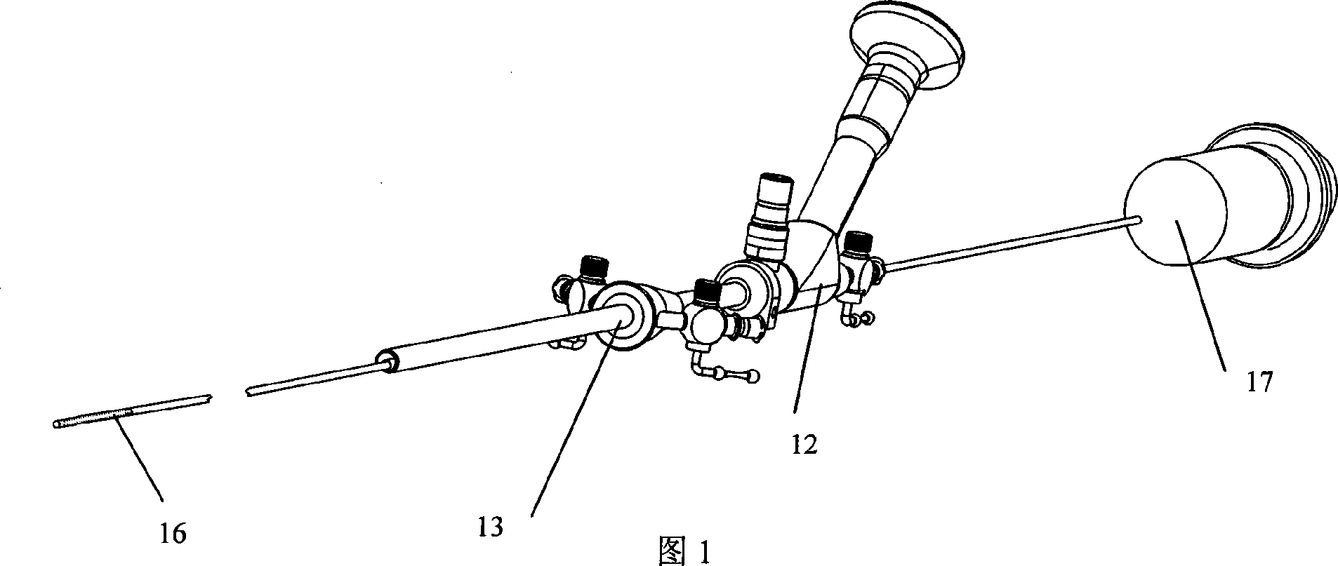
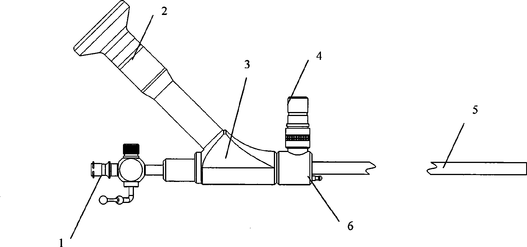
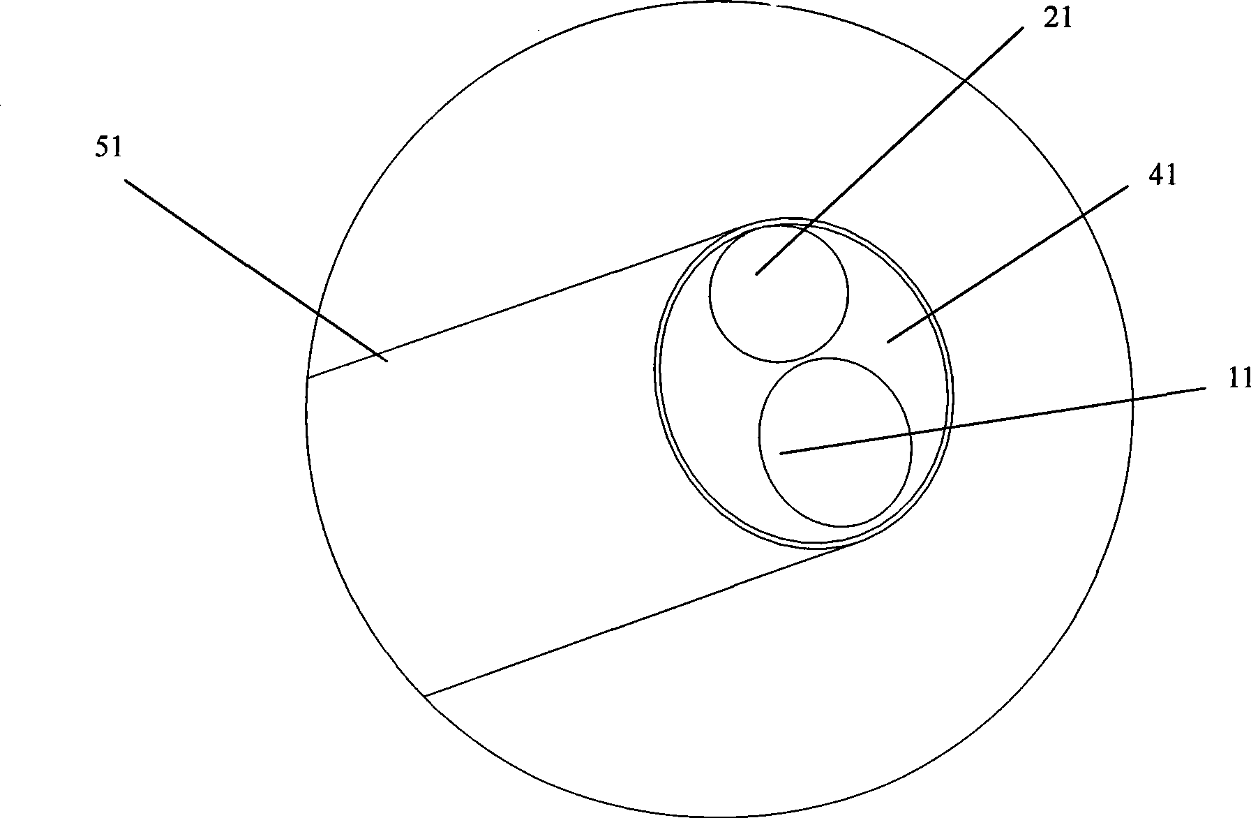
[0030] As shown in Figures 1 to 3, the ultrasonic hysteroscope system according to the present invention includes a three-channel hysteroscope with a sheath tube consisting of a double channel of the sheath tube and a single channel of the mirror body, and a three-channel hysteroscope connected to the three-channel hysteroscope. The miniature ultrasonic probe 17 of the three-channel hysteroscope includes a hysteroscope 12, a hysteroscope sheath 13 connected to the hysteroscope 12, and the hysteroscope 12 includes an endoscope main body 3 and an endoscope end 5. The instrument channel 1 connected to the end of the endoscope main body, the eyepiece input end 2 and the cold light source input end 4 arranged at an angle to the endoscope main body 3, and the connecting shaft 6 for connecting the hysteroscope sheath tube 13, and Including the instrument channel 1 passing th...


PUM

PropertyMeasurementUnit
Lengthaaaaaaaaaa
Outer diameteraaaaaaaaaa
Lengthaaaaaaaaaa

Abstract

The invention belongs to medical appliance field and in particular discloses an ultrasonic hysteroscope system. The ultrasonic hysteroscope system comprises a three-channel hysteroscope which is provided with a sheathing canal and is composed of a sheathing canal twin channels and a lens single channel, and a minisize ultrasonic probe connected with the three-channel hysteroscope. The hysteroscope is composed of an inner lens main body, an inner lens end, an appliance channel connected at the inner lens end, an ocular input end and a cold light source input end which are arranged at angles with the inner lens main body, and a connecting shaft used to connect the sheathing canal of the hysteroscope. The minisize ultrasonic probe passes through the appliance channel and the sheathing canal of the hysteroscope to detect the interior condition of the uterine cavity. The ultrasonic hysteroscope system integrates a large-diameter appliance channel in the hysteroscope, so standard appliances can enter the uterine cavity from the appliance channel of the hysteroscope, thus greatly widening operation scope. In addition, the minisize ultrasonic probe enters the uterine cavity for ultrasonic scanning from the appliance channel of the hysteroscope, so a doctor can know the pathological changes of a patient more visually and more accurately under the help of a high-resolution contrast picture and can make the best diagnosis. The ultrasonic hysteroscope system makes gynecological examination and operation methods richer and improves operation safety.

Description

technical field [0001] The invention belongs to the field of medical devices, and is a core medical tool for carrying out gynecological endoscopic examination and operation in modern medicine, in particular an ultrasonic hysteroscope system. Background technique [0002] Hysteroscopy is a minimally invasive gynecological diagnostic device used for diagnosis, treatment and follow-up of uterine cavity lesions. Hysteroscopy can not only determine the location, size, appearance and scope of the lesion, but also carefully observe the tissue structure on the surface of the lesion, and take materials or locate the curettage under direct vision, which greatly improves the diagnosis of intrauterine diseases. Accuracy, update, development and make up for the deficiencies of traditional diagnosis and treatment methods. For most patients who are suitable for diagnostic dilatation and curettage, it is more reasonable and effective to perform biopsy or dilatation and curettage after perf...

Claims


Application Information

Patent Timeline
IPC IPC(8): A61B8/12
Inventor 乔铁张阳德
Owner GUANGZHOU BAODAN MEDICAL INSTR TECH
Why Eureka
Try Eureka