Method for implementing cerebrovascular image recognition by using fast boundary tracking
A boundary tracking and image recognition technology, applied in the field of medical image recognition, can solve the problems of low boundary accuracy, insufficient smooth boundary, unsatisfactory processing speed, etc., and achieve the effect of reducing time complexity, improving accuracy and high precision
- Summary
- Abstract
- Description
- Claims
- Application Information
AI Technical Summary
Problems solved by technology
Method used
Image
Examples
Embodiment
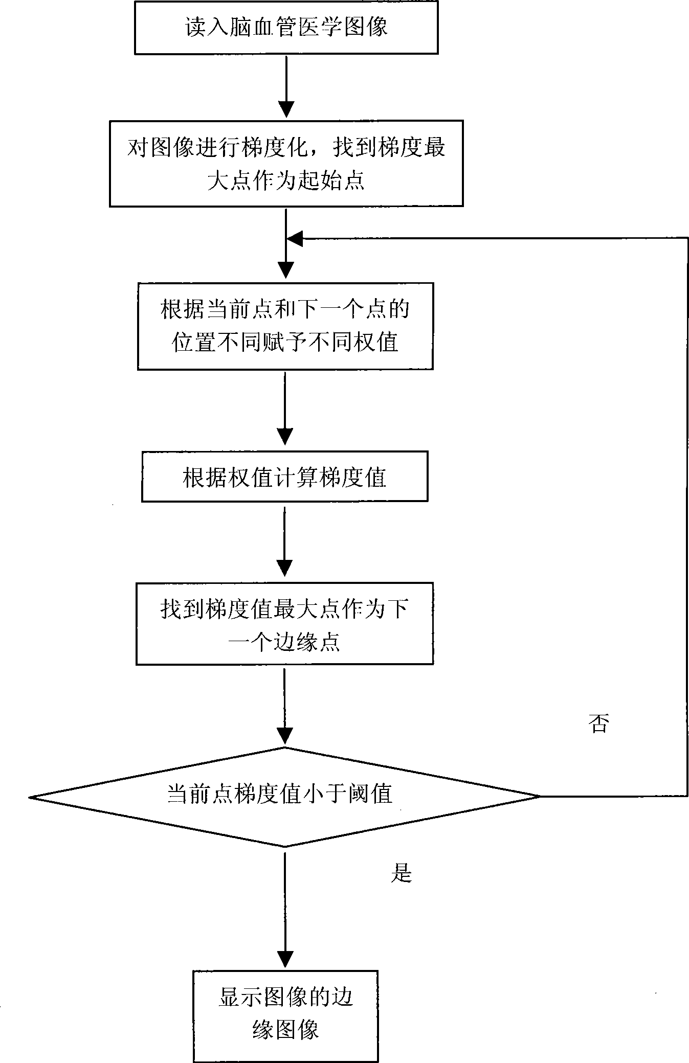
[0035] Example: figure 1 It is a flow chart of the fast boundary tracking method based on cerebrovascular image features implemented by the present invention, and the data file (picture file) is a cerebrovascular picture conforming to the BMP format.
[0036] Methods as below:
[0037] (1) Acquisition of cerebrovascular images According to the requirements of edge extraction, the DSA image files captured by the hardware equipment imaging device are read through the file reading module, and converted into standard BMP images through the image conversion module. In this embodiment, a single cerebrovascular picture is taken, and the resolution of the single picture is 1024×1024. Finally, the necessary preprocessing work is performed on the cerebrovascular images, including smoothing and denoising;
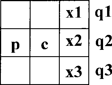
[0038] (2) Gradientization of Cerebral Vascular Images The first-order reciprocal of a digital image is based on various approximations of two-dimensional gradients. The gradient o...
PUM
 Abstract
Description
Claims
Application Information
 - R&D
- Intellectual Property
- Life Sciences
- Materials
- Tech Scout
- Unparalleled Data Quality
- Higher Quality Content
- 60% Fewer Hallucinations
Browse by: Latest US Patents, China's latest patents, Technical Efficacy Thesaurus, Application Domain, Technology Topic, Popular Technical Reports.
© 2025 PatSnap. All rights reserved.Legal|Privacy policy|Modern Slavery Act Transparency Statement|Sitemap|About US| Contact US: help@patsnap.com


