Image processing device for medical use and image processing method for medical use

An image processing device and image processing technology, applied in the directions of image data processing, image data processing, image analysis, etc., can solve the problems of decreased detection accuracy and increased burden of polyps and other lesions

Active Publication Date: 2009-04-08
OLYMPUS CORP
View PDF2 Cites 13 Cited by
  • Summary
  • Abstract
  • Description
  • Claims
  • Application Information

AI Technical Summary

Problems solved by technology

Therefore, the method for detecting lesion sites such as polyps proposed in US Patent Application No. 20030223627 is applied, for example, to three-dimensional In the case of the model, the detection accuracy of lesion sites such as

Method used


Image

  • Image processing device for medical use and image processing method for medical use
  • Image processing device for medical use and image processing method for medical use
  • Image processing device for medical use and image processing method for medical use

Examples


no. 1 Embodiment approach

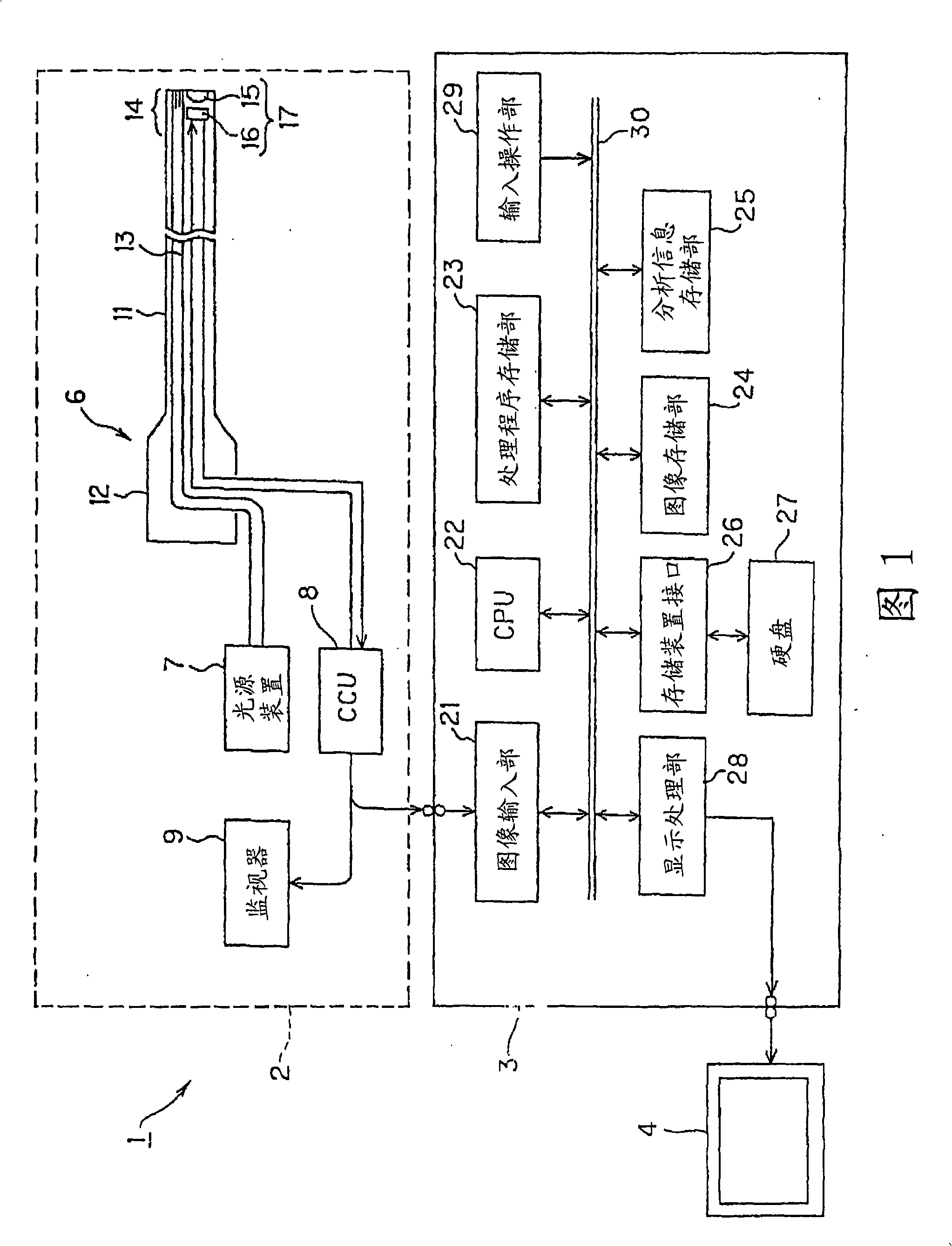
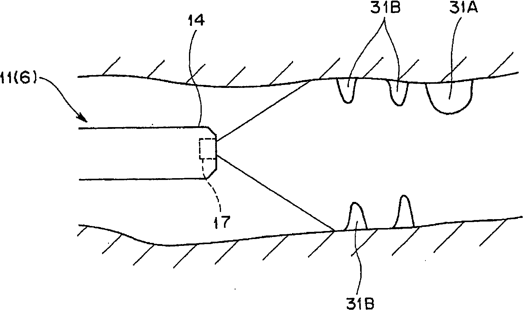
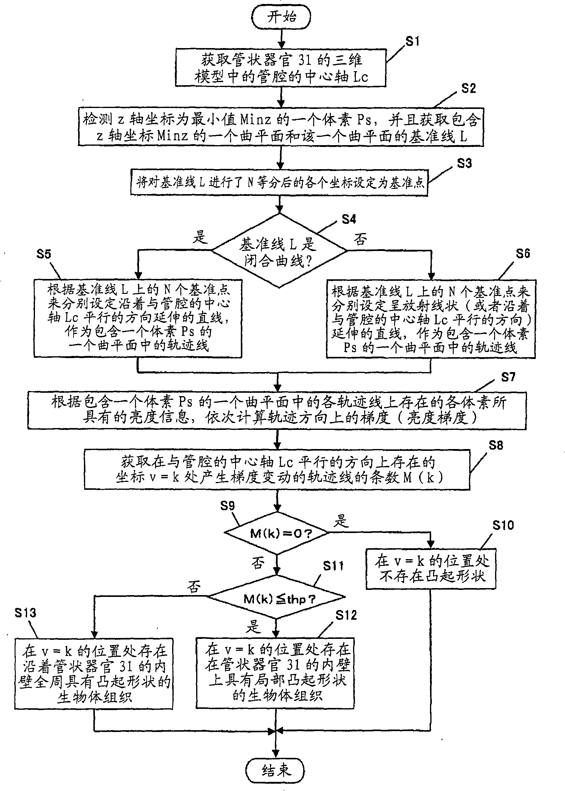
[0036] Figure 1 to Figure 7 It relates to the first embodiment of the present invention. FIG. 1 is a diagram showing an example of an overall configuration of an endoscope system using a medical image processing apparatus according to an embodiment of the present invention. figure 2 It is a schematic diagram showing a state when the endoscope of FIG. 1 is inserted into a tubular organ. image 3 It is a flowchart showing the procedure of processing performed by the medical image processing apparatus in FIG. 1 in the first embodiment. Figure 4 It is a diagram showing an example of a three-dimensional model of a living tissue estimated by the medical image processing apparatus of FIG. 1 . Figure 5 is the three-dimensional model of the living tissue estimated by the medical image processing device in FIG. Figure 4 Diagram of different examples. Figure 6 is showing Figure 4 A graph of the gradient change of each trajectory line in the 3D model of . Figure 7 is showing ...

no. 2 Embodiment approach

[0071] Figure 8 to Figure 10 It relates to the second embodiment of the present invention. In addition, detailed description of parts having the same configuration as that of the first embodiment will be omitted. In addition, the same reference numerals are used for the same constituent elements as those of the first embodiment, and description thereof will be omitted. Furthermore, the configuration of the endoscope system 1 used in this embodiment is the same as that of the first embodiment.

[0072] Figure 8 It is a flowchart showing the procedure of processing performed by the medical image processing apparatus in FIG. 1 in the second embodiment. Figure 9 is a cross-sectional view schematically showing a three-dimensional model of one living tissue estimated by the medical image processing apparatus in FIG. 1 . Figure 10 is showing Figure 9 A plot of the normal and inverse normal vectors in .

[0073] First, the CPU 22 as an edge extraction unit applies a bandpas...


PUM


Abstract

A medical image processing apparatus of the invention comprises: a three-dimensional model estimating section that estimates a three-dimensional model of body tissues based on a two-dimensional image of an image of the body tissues inside a body cavity inputted from a medical image pickup apparatus; a voxel detecting section that detects one voxel existing on a nearest side in a view direction of the medical image pickup apparatus among the voxels included in the three-dimensional model; and a raised shape detecting section that acquires one curved surface including the one voxel to detect a position with gradient variation on the one curved surface and to determine whether or not body tissues with a locally raised shape exist in the position in the three-dimensional model.

Description

technical field [0001] The present invention relates to a medical image processing device and a medical image processing method, and more particularly to a medical image processing device and a medical image processing method capable of detecting convex shapes in a three-dimensional model of living tissue in a body cavity. Background technique [0002] An endoscope system including an endoscope and a medical image processing device is widely used in the medical field and the like. Specifically, the endoscope system is configured to include, for example: an endoscope configured to have an insertion portion for being inserted into a body cavity of a living body, an objective optical system provided at a distal end portion of the insertion portion, and an endoscope for An imaging unit that captures an image in a body cavity formed by the objective optical system and outputs it as an imaging signal; The image shows the processing of the image inside the body cavity. Furthermor...

Claims


Application Information

Patent Timeline
IPC IPC(8): A61B1/04G06T1/00
CPCG06T7/0083G06T2207/10016A61B1/05G06T17/30G06T2207/30032G06T2207/10068G06T7/0065G06T2207/30028G06T7/0012G06T7/12G06T7/55
Inventor 井上凉子西村博一田中秀树中村健次沢美穗
Owner OLYMPUS CORP
Why Eureka
Try Eureka