
Method for enhancing blood vessels in retinal images based on the directional field

A retina and direction field technology, applied in the field of image processing, can solve problems such as difficulties in automatic realization

Inactive Publication Date: 2009-09-02
INST OF AUTOMATION CHINESE ACAD OF SCI
View PDF0 Cites 20 Cited by
  • Summary
  • Abstract
  • Description
  • Claims
  • Application Information

AI Technical Summary

Problems solved by technology

[0003] Retinal vessel segmentation is very important, but its automatic implementation is quite difficult

Method used


Image

  • Method for enhancing blood vessels in retinal images based on the directional field
  • Method for enhancing blood vessels in retinal images based on the directional field
  • Method for enhancing blood vessels in retinal images based on the directional field

Examples


Embodiment Construction

[0039] In order to make the object, technical solution and advantages of the present invention clearer, the present invention will be described in further detail below in conjunction with specific embodiments and with reference to the accompanying drawings.

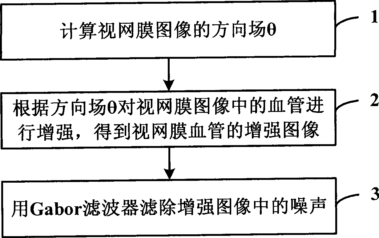
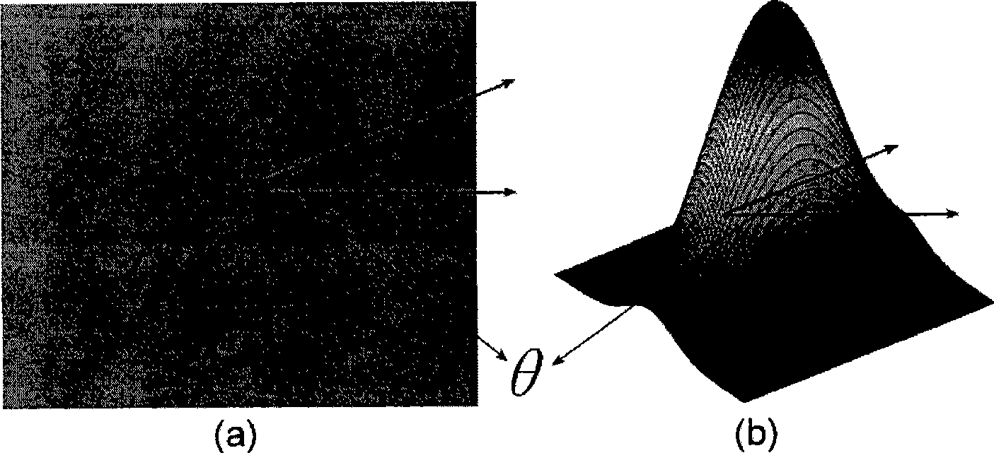
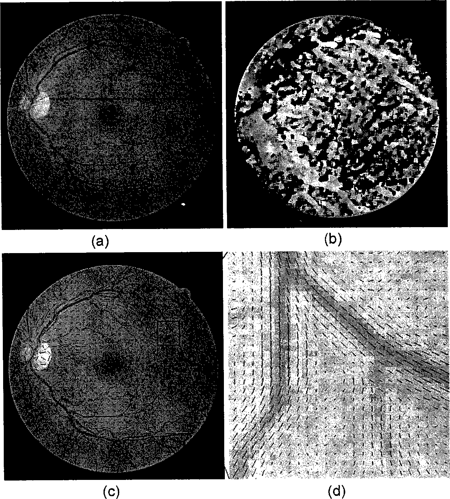
[0040] The core idea of ​​the present invention is to propose a method for enhancing retinal image blood vessels based on a direction field, and to enhance the image point by point under the guidance of a fine direction field. The method includes the following steps: Estimating the direction field θ of the retinal image; automatically enhancing the blood vessels in the image under the guidance of the direction field; filtering out the noise in the enhanced image with a Gabor filter.

[0041] like figure 1 as shown, figure 1 A flow chart of the method for enhancing retinal image blood vessels based on the direction field provided by the present invention, the method includes the following steps:

[0042] Step 1: Calculat...


PUM


Abstract

The invention discloses a method for enhancing blood vessels in retinal images based on the directional field, which includes: step one: the directional field Theta of a retinal image is calculated; step two: the blood vessels in the retinal image are enhanced according to the directional field Theta to obtain the enhanced image of retinal blood vessels; and step three: a Gabor wave filter is used for filtering noise in the enhanced image. The method can effectively enhance small blood vessels with low contrast ratio in the retinal images, which brings great convenience to the subsequent processing of the retinal images. In the invention, normalization processing is carried out to image backgrounds while the blood vessels are enhanced, which effectively overcomes influence of image noise and uneven lightness and saves the single normalization step. The invention has important practical value in the fields of dissection of blood vessels in the retinal and heart images and other fields.

Description

technical field [0001] The invention relates to image processing technology, in particular to a method for enhancing retinal image blood vessels based on a direction field. Background technique [0002] With the development of imaging technology, in the application of ophthalmology, fundus photography technology is more and more widely used. This is not only because of the in-depth application of computer technology, but also because fundus images can reflect the lesions of many diseases in a timely manner. The retina is the only organ in the human body that can directly obtain image information of blood vessels in vivo. These blood vessel information play a vital role in the diagnosis and treatment of many diseases. The segmentation of the retinal vascular network is very important for extracting these vascular information, and the results can be used to locate landmarks for image registration; it can be used to estimate the difference between arterial blood flow and venou...

Claims


Application Information

Patent Timeline
IPC IPC(8): G06T5/00G06T5/10A61B3/14
Inventor 田捷陈健杨鑫
Owner INST OF AUTOMATION CHINESE ACAD OF SCI
Learn More