Method for processing medical images

A processing method and medical image technology, applied in the field of medical image processing, can solve problems such as inability to give accurate 3D images, difficult treatment, distortion, etc.

Inactive Publication Date: 2010-01-13
SHENZHEN INST OF ADVANCED TECH
View PDF0 Cites 29 Cited by
  • Summary
  • Abstract
  • Description
  • Claims
  • Application Information

AI Technical Summary

Problems solved by technology

However, the obtained slices can only provide two-dimensional images of the inside of the human body, and the two-dimensional images can only represent the anatomical information of a certain interface. Doctors can only estimate the size and shape of the lesion from multiple two-dimensional images based on experience. "The three-dimensional geometric relationship between the lesion and its surrounding tissue, which brings difficulties to the treatment
In addition, in radiotherapy applications, simple coordinate superposition of some anatomical information on two-dimensional images cannot give accurate three-dimensional images, which may easily cause distortion and distortion of lesion positioning

Method used


Image

  • Method for processing medical images
  • Method for processing medical images
  • Method for processing medical images

Examples


Embodiment Construction

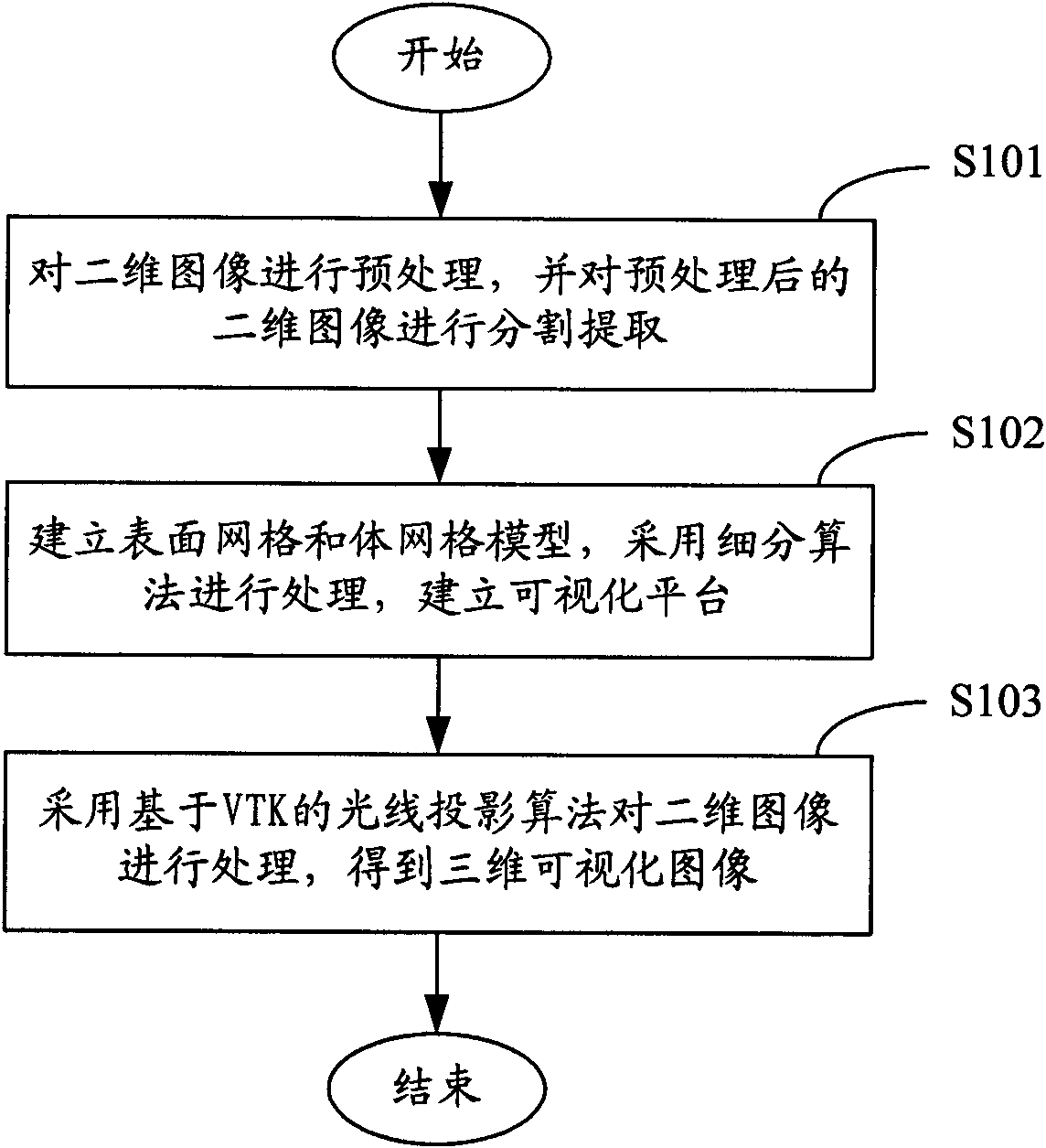
[0023] figure 1 The medical image processing method flow in one embodiment is shown, and the specific process is as follows:

[0024] In step S101, the two-dimensional image is preprocessed, and the preprocessed two-dimensional image is segmented and extracted.

[0025] In step S102, a surface mesh and a volume mesh model are established, processed with a subdivision algorithm, and a visualization platform is established.

[0026] In step S103, a VTK-based ray projection algorithm is used to process the two-dimensional image to obtain a three-dimensional visualized image.

[0027] The aforementioned preprocessing of the two-dimensional image includes processing such as image sharpening and denoising. In one embodiment, the preprocessed two-dimensional image is segmented and extracted by using an automatic segmentation algorithm and a manual segmentation. For different segmentation objects (such as outer contour, internal structure or other special parts), it is necessary to...


PUM


Abstract

The invention provides a method for processing medical images, comprising the following steps: A. c preprocessing two-dimensional images, and partitioning and extracting the preprocessed two-dimensional images; B. establishing surface mesh and body mesh moulds, adopting a subdivision algorithm for processing, and establishing a visualization platform; and C. adopting a light projection algorithm based on VTK for processing the two-dimensional images, and obtaining three-dimensional visualization images. When the method for processing medical images provided by the invention is adopted, fast batch processing can be carried out on the medical two-dimensional images, and three-dimensional visualization of the two-dimensional images can be realized.

Description

technical field [0001] The present invention relates to the technical field of image processing, and more specifically, to a medical image processing method. Background technique [0002] Computed Tomography (CT for short) and Magnetic Resonance Imaging (MRI for short) are commonly used imaging techniques in medical imaging diagnosis. Slice images to find diseased variants. However, the obtained slices can only provide two-dimensional images of the inside of the human body, and the two-dimensional images can only represent the anatomical information of a certain interface. Doctors can only estimate the size and shape of the lesion from multiple two-dimensional images based on experience. "The three-dimensional geometric relationship between the lesion and its surrounding tissue is revealed, which brings difficulties to the treatment. In addition, in radiotherapy applications, simple coordinate superposition of certain anatomical information on two-dimensional images cannot...

Claims


Application Information

Patent Timeline
Patent Type & Authority Applications(China)
IPC IPC(8): G06T15/00G06T15/50G06T17/00G06T7/00
Inventor 胡战利郑海荣
Owner SHENZHEN INST OF ADVANCED TECH
Why Eureka
Try Eureka