Computer aided detection method for microcalcification in mammograms

A technology of micro-calcification and calcification points, which is applied in the field of automatic analysis and processing of medical images, can solve the problems of high false positive rate of detection results and difficulty in detecting micro-calcification points completely and correctly, so as to improve the degree of automation, reduce workload, improve The effect of detection speed

Inactive Publication Date: 2010-10-06
XIAN UNIV OF TECH
View PDF2 Cites 29 Cited by
  • Summary
  • Abstract
  • Description
  • Claims
  • Application Information

AI Technical Summary

Problems solved by technology

[0005] The purpose of the present invention is to provide a computer-aided detection method for breast microcalcifications, which solves the problems in the prior art that it is difficult to completely and correctly detect microcalcifications in dense mammograms and the false positive rate of detection results is too high question

Method used


Image

  • Computer aided detection method for microcalcification in mammograms
  • Computer aided detection method for microcalcification in mammograms
  • Computer aided detection method for microcalcification in mammograms

Examples


Embodiment 1

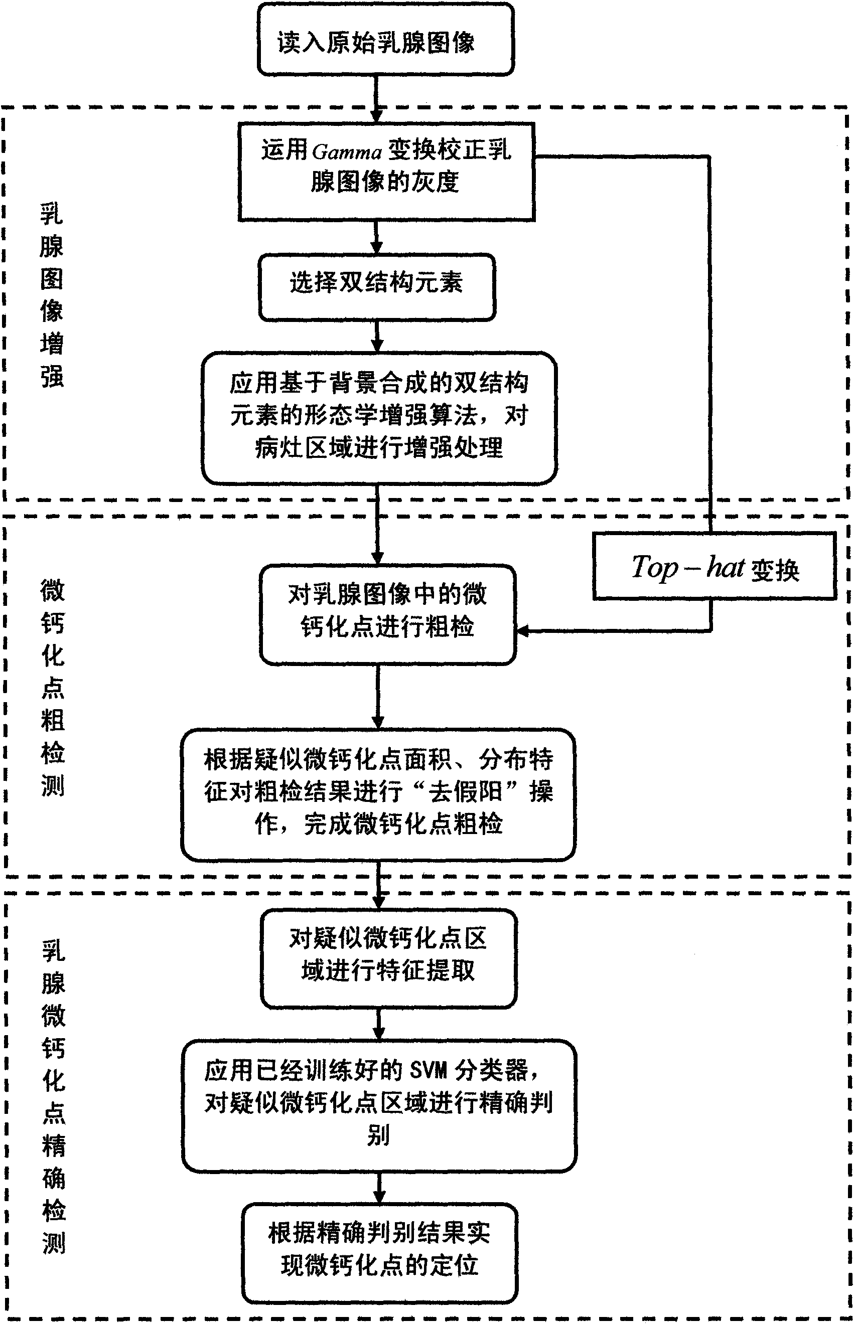
[0051] A method for computer-aided detection of mammary gland microcalcifications is implemented through the following steps:

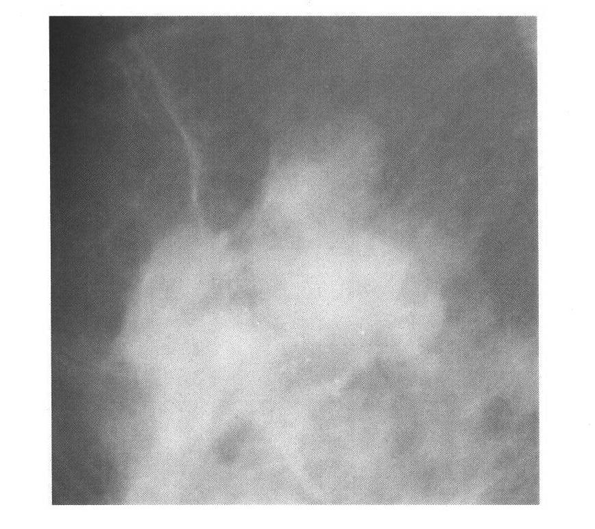
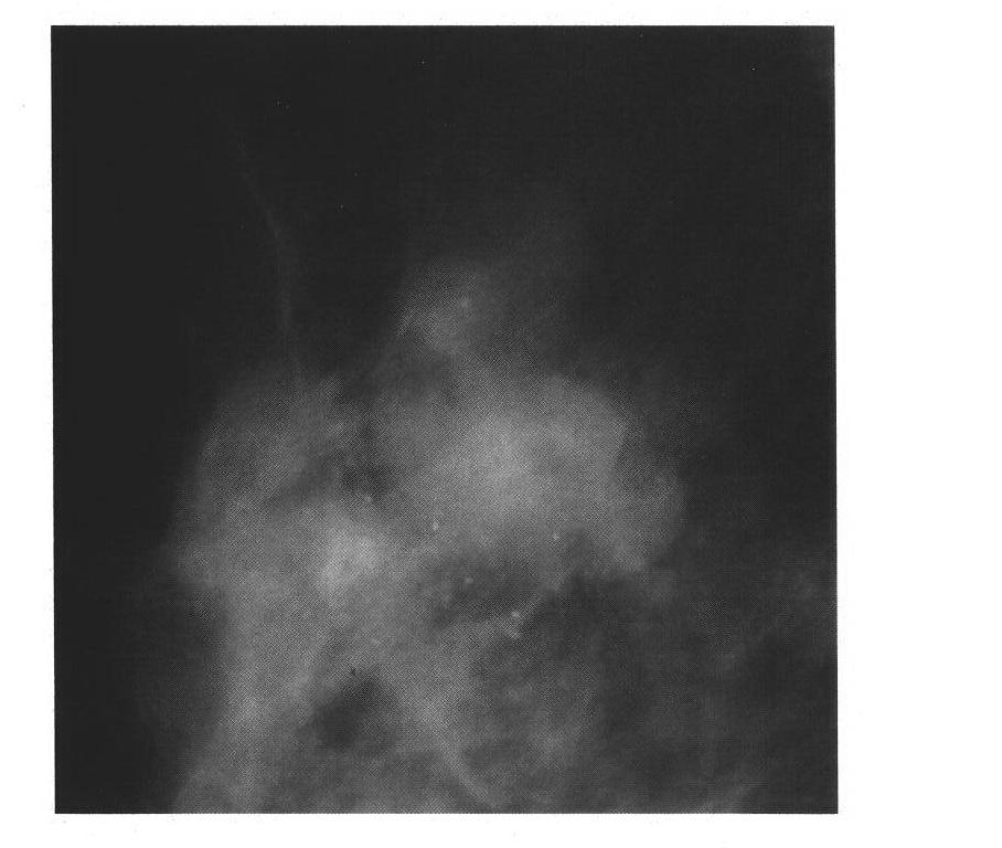
[0052] Step 1, read a picture such as figure 2 The original mammary gland image shown, and then use the Gamma grayscale correction method to perform grayscale correction on the original mammary gland image:

[0053] The Gamma grayscale correction method is: Among them, I(x, y) is the input original image, F(x, y) is the image after Gamma grayscale correction, and the value of γ is 3, and the image after grayscale correction is obtained as image 3 shown;

[0054] Step 2, using the background overlay method based on double structural elements to enhance image 3 Details of the center position and edge area of ​​the target area of ​​suspected microcalcifications in:

[0055] Using double structuring elements: and right image 3 Perform the following morphological processing to obtain the image G 1 (x, y) and G 2 (x,y):

[0056] ...


PUM


Abstract

The invention discloses a computer aided detection method for microcalcification in mammograms, which comprises the following steps of: performing grayscale correction and transformation on a mammogram image to obtain an image after grayscale correction; acquiring microcalcification enhanced mammogram image by adopting dual-structure element-based background superposition method, and simultaneously acquiring another background suppressed mammogram image by adopting a Top-hat transformation method in morphology; segmenting the two images by adopting double thresholds to acquire a preliminary microcalcification image, and performing postprocessing to form a coarse microcalcification detection image; and extracting characteristics from coarsely detected microcalcification destination area, classifying by using a support vector machine, removing false microcalcification destination areas, marking the rest microcalcification into the mammogram image for the reading of doctors. The method can free the doctors from trivial mammogram reading and classifying work, and assists the doctors in better understanding and judging the images so as to reduce error diagnosis and missed diagnosis and fulfill the aim of improving diagnostic accuracy.

Description

technical field [0001] The invention belongs to the technical field of automatic analysis and processing of medical images, and in particular relates to a computer-aided detection method for breast microcalcification points. Background technique [0002] Breast cancer is one of the most common malignant tumors in women all over the world, which seriously threatens women's health and even life. In developed countries such as Northern Europe and the West, the incidence of breast cancer ranks first in the incidence of female malignant tumors. In my country, with the change of people's living and eating habits, the incidence of breast cancer has also shown an obvious upward trend, and the incidence of breast cancer among women aged 25-34 has also increased rapidly. In recent decades, with the development of medical technology, the diagnosis and treatment of breast cancer have made great progress. However, the mortality rate of breast cancer has not decreased significantly, whic...

Claims


Application Information

Patent Timeline
Patent Type & Authority Applications(China)
IPC IPC(8): G06K9/00G06T5/00G06T7/00G06T7/136
Inventor 张二虎王帆
Owner XIAN UNIV OF TECH
Why Eureka
Try Eureka