Eureka AIR delivers breakthrough ideas for toughest innovation challenges, trusted by R&D personnel around the world.

Agarose gel micro-flow chamber device for detecting cell migration

A technology of agarose gel and cell migration, applied in measuring devices, enzymology/microbiology devices, flow characteristics, etc., can solve problems such as poor sealing, experimental failure, leakage, etc., and achieve good sealing effect

Inactive Publication Date: 2012-01-11
CHONGQING MEDICAL UNIVERSITY
View PDF0 Cites 0 Cited by
  • Summary
  • Abstract
  • Description
  • Claims
  • Application Information

AI Technical Summary

Problems solved by technology

However, the non-bonding method of this device leads to its poor sealing, and frequent liquid leakage leads to the failure of the experiment.
In addition, the device cannot adjust its liquid flow rate through a micro-injection pump

Method used


Image

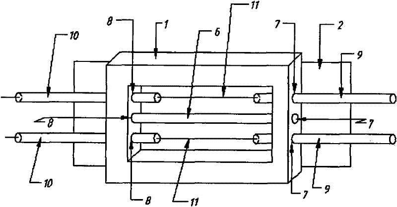
  • Agarose gel micro-flow chamber device for detecting cell migration
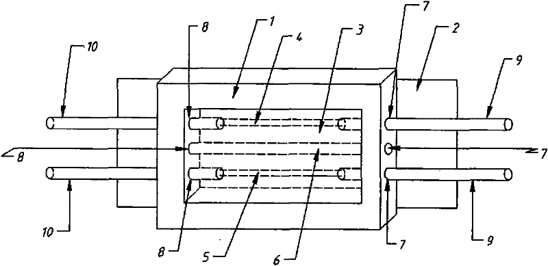
  • Agarose gel micro-flow chamber device for detecting cell migration
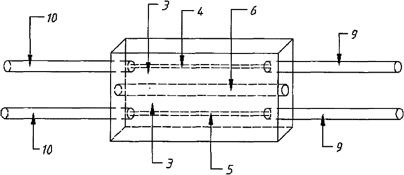
  • Agarose gel micro-flow chamber device for detecting cell migration

Examples


Embodiment

[0016] 1.1 Main materials

[0017] Human endothelial cell line EA.hy926, purchased from Cell Resource Center, Shanghai Institutes for Biological Sciences, Chinese Academy of Sciences; DMEM F12 medium, purchased from Invitrogen (USA); fetal bovine serum, purchased from Hangzhou Sijiqing Company; chemokine; agar Glycogel was purchased from Advancebiomatrx (USA).

[0018] 1.2 Cell Culture

[0019] Cells were grown adherently in 10% fetal bovine serum at 37°C, 5% CO 2 Cultivated in an incubator.

[0020] 1.3 Fabrication of microfluidic device

[0021] Arranging the microfilaments: laser perforation was carried out on the front and rear two side walls of the mouth-shaped plexiglass frame 1 with an inner diameter of 6 cm long, a width of 3 cm, and a thickness of 1 cm to obtain 3 inlet ports 7 and 8 outlets each with a diameter of 1 mm, 4 Teflon thin tubes with a diameter of 1mm are inserted into both sides of the inlet 7 and the outlet 8 one by one, wherein the inlet pipe 9 is c...


PUM

PropertyMeasurementUnit
widthaaaaaaaaaa
lengthaaaaaaaaaa
pore sizeaaaaaaaaaa

Abstract

The invention relates to an agarose gel micro-flow chamber device for detecting cell migration. The device comprises a square organic glass frame (1) and a glass slide (2) padded below the square organic glass frame, wherein the square organic glass frame (1) is filled with agarose gel (3); three parallel micro-channels in the same depth pass through the agarose gel (3); the two side walls of thesquare organic glass frame (1) are provided with a lead-in port (7) and a lead-out port (8); and a liquid inlet pipe (9) and a liquid outlet pipe (10) are inserted into the gel through the lead-in port (7) and the lead-out port (8) and connected with the micro-channels (4 and 5) on the two sides. The device has the advantages of simple preparation process, high tightness, capability of producing stable concentration gradient in a short time and wide application in various cell migration tests.

Description

technical field [0001] The invention relates to the field of biochips, in particular to an agarose gel microfluidic chamber device for detecting cell migration. Background technique [0002] Cell migration plays an important role in many physiological and pathological processes. The traditional cell migration detection device includes Boyden chamber and Transwell chamber. The technical solution is: Boyden chamber and Transwell chamber are a small box device, and the box is divided into upper and lower boxes by a microporous filter membrane with a 3-5 μm pore size. Two chambers, the upper chamber was added with the suspension of the tested cells; the lower chamber was added with chemokine, and incubated at 37°C for several hours, the cells in the upper chamber were induced by the chemokines in the lower chamber, and the cells were allowed to pass through the filter membrane micropores. Enter the filter membrane, and finally take the filter membrane. After the steps of fixati...

Claims


Application Information

Patent Timeline
Patent Type & Authority Patents(China)
IPC IPC(8): C12M1/34G01N11/10
Inventor 熊新
Owner CHONGQING MEDICAL UNIVERSITY
Why Eureka
Eureka Blog
Learn More