Three-dimensional reconstruction method for two-dimensional ultrasonic image

A technology of 3D reconstruction and ultrasound image, which is applied in the field of medical image processing, can solve the problems of poor quality and slow reconstruction speed of 3D image reconstruction, and achieve the effect of improving reconstruction speed, strong sense of reality, and clear reconstruction image

Inactive Publication Date: 2011-06-29
PHILIPS CHINA INVESTMENT
View PDF0 Cites 36 Cited by
  • Summary
  • Abstract
  • Description
  • Claims
  • Application Information

AI Technical Summary

Problems solved by technology

[0009] Aiming at the shortcomings of the above-mentioned poor quality of three-dimensional image reconstruction and slow reconstruction speed in the prior art, the technical problem to

Method used


Image

  • Three-dimensional reconstruction method for two-dimensional ultrasonic image
  • Three-dimensional reconstruction method for two-dimensional ultrasonic image
  • Three-dimensional reconstruction method for two-dimensional ultrasonic image

Examples


Embodiment 1

[0057] The present invention proposes a technical scheme for three-dimensional reconstruction of the collected two-dimensional sequence image data, which can achieve According to the requirement of clinical application, at the same time, the synthetic image noise is processed to better display the detailed information of the 3D reconstructed image.

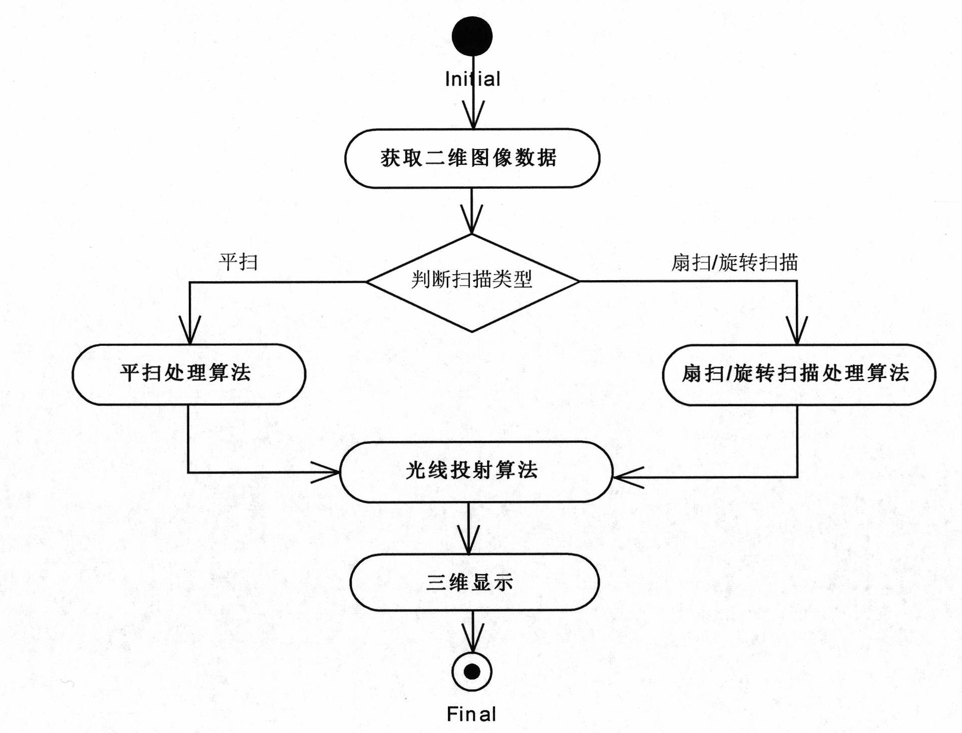
[0058] Such as figure 2 As shown, the three-dimensional reconstruction method of the two-dimensional ultrasonic image of the present invention comprises the following steps:

[0059] Obtain two-dimensional image data through mechanical drive scanning;

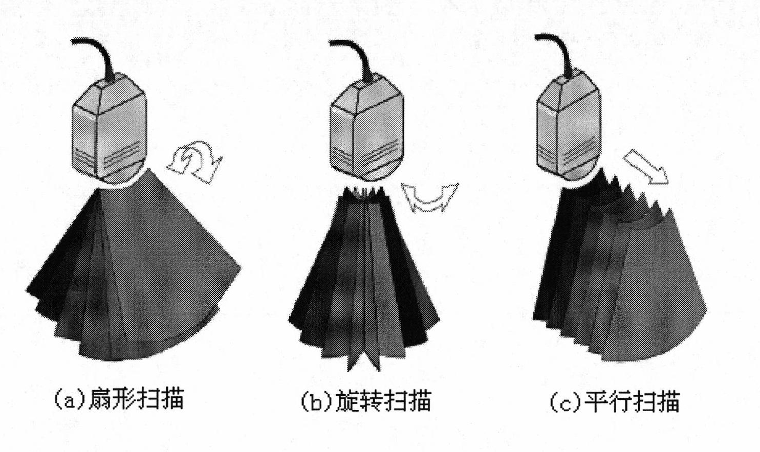
[0060] Determine the scan type, if it is a sector scan, use the sector scan processing algorithm to process the two-dimensional image data to generate a regular volume data field;

[0061] The above regular volume data field is used to display a three-dimensional image through a ray casting algorithm based on space volume rendering.

[0062] The present invention uses a mechani...

Embodiment 2

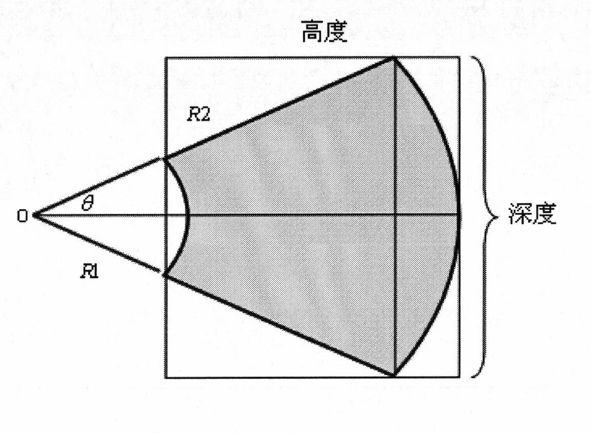
[0087] The difference from Embodiment 1 is that if the scan type is judged to be rotary scanning, then the two-dimensional image data is processed using a rotary scanning processing algorithm to generate a regular volume data field, and the steps include extracting the region of interest and calculating the regular volume data The size of the field, the improved coordinate conversion and interpolation algorithm, and the filtering process. The size of the regular body data field is different from the sector scanning method, specifically:

[0088] Such as Figure 4 As shown in , it is stipulated that the width of the regular volume data field is the width of the ROI data, and the height and depth of the regular volume data field are both twice the height of the ROI data.

Embodiment 3

[0090] The difference with Embodiment 1 or 2 is that: if the scan type is judged to be parallel scan, a parallel scan processing algorithm is used to process the two-dimensional image data to generate a regular volume data field, and the steps include: extracting the region of interest, calculating The size of the regular volume data field, the conversion of the pixel point data in the 2D image to the 3D volume data, and the filtering process. The calculation of the size of the regular volume data field is different from the sector scanning and rotation scanning methods, specifically: specifying the region of interest The width and height of the data are the width and height of the regular body data field, and the depth of the regular body data field, that is, the length in the Z direction, is taken as the ratio of the scanning length of the plain scan to the scanning time;

[0091] Such as Figure 12 As shown, the data of the region of interest is placed in the regular volume...


PUM


Abstract

The invention relates to a three-dimensional reconstruction method for a two-dimensional ultrasonic image. The method comprises the following steps of: acquiring data of a two-dimensional image in a mechanically-driven scanning mode; judging a scanning type, and processing the data of the two-dimensional image by adopting a sector scanning/rotary scanning processing algorithm to generate a regular data field if the scanning is sector scanning or rotary scanning; and displaying a three-dimensional image by utilizing the regular data field through a ray casting algorithm. The sector scanning processing algorithm comprises the processing steps of: extracting interesting areas, namely rejecting unwanted data information; calculating the size of the regular data field to obtain the length, width and height of the regular data field; improving coordinate conversion and an interpolation algorithm to obtain a grey value of a current pixel; and performing filtering processing. In the method, the three-dimensional reconstruction is performed on the data of the two-dimensional ultrasonic image which are acquired in different scanning modes; and the method has the advantages of clear reconstructed images, strong sense of reality, high imaging speed and the like.

Description

technical field [0001] The invention relates to medical image processing technology, in particular to a three-dimensional reconstruction method of two-dimensional ultrasonic images. Background technique [0002] Compared with other technologies, ultrasonic medicine has obvious advantages. Because it is non-invasive, it has the characteristics of harmless, painless, and intuitive use. It is one of the indispensable imaging diagnostic methods in clinical medicine and has been widely used in clinical medicine. application. Traditional B-mode ultrasound imaging systems can only provide two-dimensional sequential images of scanned objects, and doctors can only reconstruct these tomographic images into three-dimensional objects through the human brain, which requires doctors to have considerable experience and spatial imagination. [0003] Three-dimensional ultrasound imaging has the advantages of intuitive image display, accurate measurement of medical diagnosis parameters, and ...

Claims


Application Information

Patent Timeline
IPC IPC(8): A61B8/00G06T7/00G06T15/00
Inventor 朱力军赵建
Owner PHILIPS CHINA INVESTMENT
Why Eureka
Try Eureka