Image-recognition-technology-based shooting-speed-adjustable wireless capsule endoscope system and method

A technology for capsule endoscopy and image recognition, which is applied to endoscopy, in vivo radio detectors, medical science, etc., can solve the problems of missed detection and false detection of photo information, so as to reduce missed detection and false detection and reduce the possibility of performance, improve accuracy

Active Publication Date: 2012-04-04
XIAN JIAOTONG LIVERPOOL UNIV
View PDF11 Cites 9 Cited by
  • Summary
  • Abstract
  • Description
  • Claims
  • Application Information

AI Technical Summary

Problems solved by technology

It solves the problems of false detection and missed detection caused by factors such as image processing mode, shooting rate, and photo information amount in the wireless capsule endoscope system in the prior art

Method used


Image

  • Image-recognition-technology-based shooting-speed-adjustable wireless capsule endoscope system and method
  • Image-recognition-technology-based shooting-speed-adjustable wireless capsule endoscope system and method
  • Image-recognition-technology-based shooting-speed-adjustable wireless capsule endoscope system and method

Examples


Embodiment

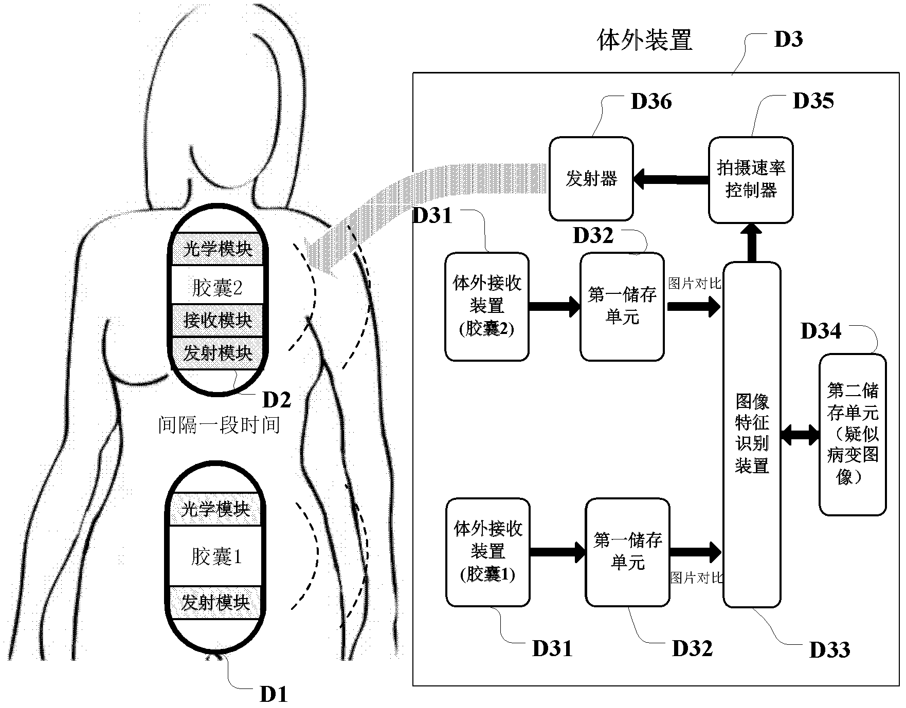
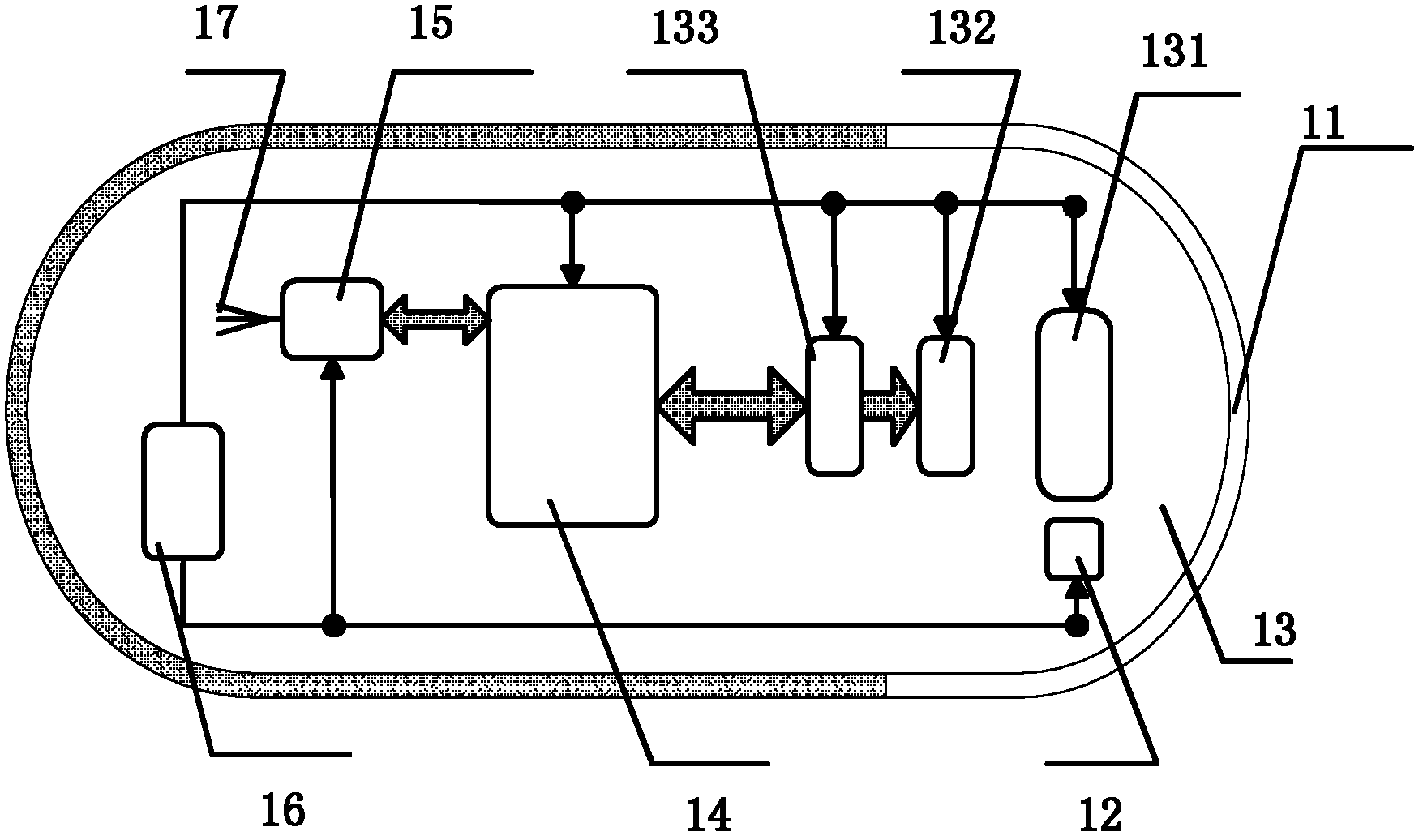
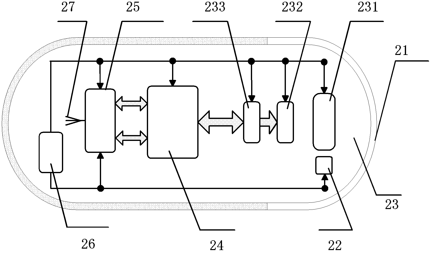
[0042] Such as figure 1 Shown is a wireless capsule endoscope system with adjustable shooting rate based on image recognition technology, including a wireless capsule endoscope and an extracorporeal device D3. The first wireless capsule endoscope D1 and the second wireless capsule endoscope D2 that adjusts its own shooting rate according to the reference image information captured by the first wireless capsule endoscope in vivo, the external device includes a wireless capsule endoscope for receiving An external receiving device D31 for the in-vivo captured images of the wireless capsule endoscope, a first storage unit D32 for storing the in-vivo captured images of the wireless capsule endoscope, and an image feature recognition device for performing image feature recognition on the stored in-vivo captured images of the wireless capsule endoscope The device D33, the second storage unit D34 for storing the in-vivo captured image of the wireless capsule endoscope as a suspected l...


PUM


Abstract

The invention discloses a wireless capsule endoscope system. A wireless capsule endoscope at least comprises a first wireless capsule endoscope and a second wireless capsule endoscope, wherein the first wireless capsule endoscope is put into a body first and shoots images which serve as reference images inside the body; and the second wireless capsule endoscope adjusts the shooting speed of the second wireless capsule endoscope according to the information of the reference images shot by the first wireless capsule endoscope inside the body. Based on a suspected diseased area, the wireless capsule endoscope system changes the shooting speed in a targeted way, so the number of pictures of the suspected diseased area is increased effectively, namely the ratio of the pictures which need more observation to the total number of the pictures is raised, the burden of post image processing and screening is lowered, and doctors can diagnose more conveniently.

Description

technical field [0001] The invention relates to a wireless capsule endoscope system and method for detecting a suspected lesion area through an existing image recognition technology and adjusting a shooting rate. Background technique [0002] The history of humans using endoscopes to detect internal organs can be traced back to the 19th century. In 1806, German scientist Philipp Bozzini first invented the prototype of an endoscope that used candlelight to detect human bladder and intestines. Since then, different types of endoscopes (such as gastroscopes, used to detect gastric lesions; colonoscopes, used to detect intestinal lesions) have been continuously developed and improved to help doctors detect diseases of the digestive system in vivo . [0003] In the past ten years, endoscope-related science and technology have developed rapidly, especially the birth of a medical auxiliary device that can be used to supplement and improve gastroscope and colonoscope—wireless capsu...

Claims


Application Information

Patent Timeline
Patent Type & Authority Applications(China)
IPC IPC(8): A61B1/04A61B1/045A61B5/07
Inventor 罗天明林永义王炤陈瑾慧
Owner XIAN JIAOTONG LIVERPOOL UNIV
Why Eureka
Try Eureka