Temperature imaging method and system

An imaging method and imaging system technology, applied in medical science, sensors, diagnostic recording/measurement, etc., can solve the problems of not meeting the real-time requirements of temperature imaging, reduced treatment effect, normal cell damage, etc., to reduce calculation errors , improve the calculation speed, and avoid the effect of unwinding

Inactive Publication Date: 2012-07-18
SHENZHEN INST OF ADVANCED TECH CHINESE ACAD OF SCI
View PDF3 Cites 10 Cited by
  • Summary
  • Abstract
  • Description
  • Claims
  • Application Information

AI Technical Summary

Problems solved by technology

However, phase warping is very common in phase maps. Unwrapping is the first step in reference-free temperature imaging based on proton resonance frequency shift. Each phase image needs to be unwrapped before temperature calculation. , and the unwinding calculation is quite time-consuming, which cannot meet the real-time requirements of temperature imaging. Especially in clinical applications, it is possible that normal cells will be damaged if the temperature is too high due to the inability to monitor the temperature in real time, and the therapeutic effect will also be affected. decrease accordingly

Method used


Image

  • Temperature imaging method and system
  • Temperature imaging method and system
  • Temperature imaging method and system

Examples


Embodiment Construction

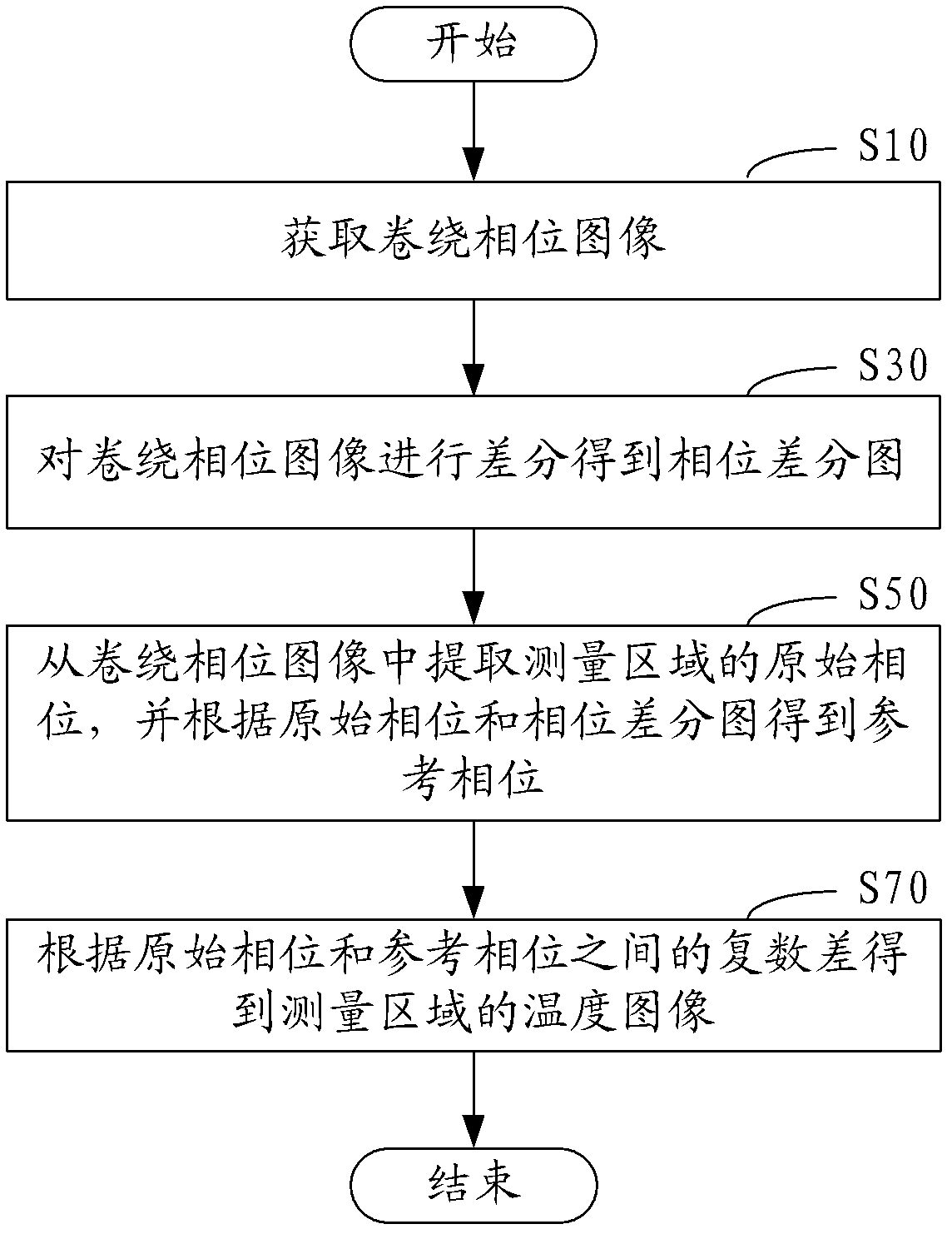
[0052] In one embodiment, such as figure 1 Shown, a kind of temperature imaging method, comprises the following steps:

[0053] Step S10, acquiring a wrapping phase image.

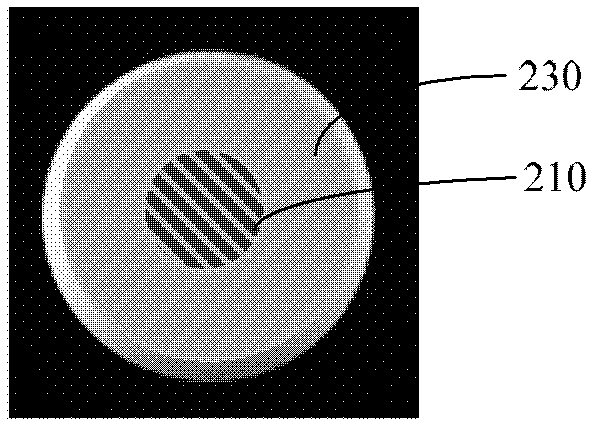
[0054] In this embodiment, the corresponding wrapping phase image can be obtained after reconstructing the data obtained in the imaging sequence. Such as figure 2 As shown, the winding phase diagram is roughly divided into two continuous areas, the measurement area 210 and the reference area 230, wherein the measurement area 210 is the corresponding heating area in the treatment of thermal ablation of tumors using high-intensity focused ultrasound, and the reference area 230 is For the unheated region, the associated phase can be obtained from the winding phase diagram.

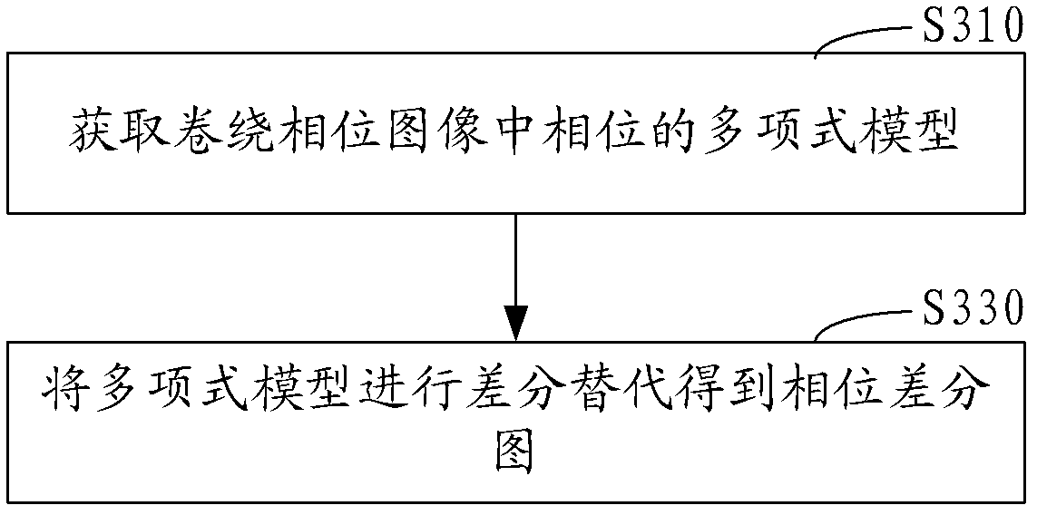
[0055] Step S30, performing a difference on the warp phase image to obtain a phase difference map.

[0056] In this embodiment, the time-consuming unwrapping of the image to obtain the phase difference map is avoided by performing di...


PUM


Abstract

The invention relates to a temperature imaging method which comprises the steps of acquiring a wrapping phase image; carrying out difference on the wrapping phase image to obtain a phase difference image; extracting an original phase of a measurement region from the wrapping phase image and obtaining a reference phase according to the original phase and the phase difference image; and obtaining a temperature image of the measurement region according to a complex difference between the original phase and the reference phase. According to the temperature imaging method and the temperature imaging system, the phase difference image is obtained by carrying out difference on the wrapping phase image, and the two-dimensional phase unwrapping is avoided, so that the computation speed is greatly improved, and the requirement of temperature imaging on the real-time property is further met on the premise of not losing the accuracy.

Description

【Technical field】 [0001] The invention relates to magnetic resonance technology, in particular to a temperature imaging method and technology. 【Background technique】 [0002] Minimally invasive hyperthermia is more and more widely used in the treatment of benign and malignant solid tumors. Its mechanism is to use high-intensity focused ultrasound to instantly increase the temperature of tumor tissue to above 60°C, which in turn leads to tumor cell damage and tissue coagulation. necrosis. In the implementation of minimally invasive hyperthermia, it is necessary to heat to above 60°C to cause tumor cell necrosis, but the heating temperature must be controlled within 100°C, otherwise the tissue will be carbonized and difficult to metabolize and absorb. In order to ensure that the tumor cells are completely killed At the same time, normal cells are protected from or less damaged. Monitoring temperature imaging has become the basic guarantee for safe treatment. Magnetic resonanc...

Claims


Application Information

Patent Timeline
Patent Type & Authority Applications(China)
IPC IPC(8): A61B5/01A61B5/055
Inventor 何梦玥邹超钟耀祖饶芳刘新郑海荣
Owner SHENZHEN INST OF ADVANCED TECH CHINESE ACAD OF SCI
Why Eureka
Try Eureka