Three-dimensional mammary gland ultrasound image partition method based on homoplasmon and partial energy

An ultrasound image, local energy technology, applied in the field of medical ultrasound image processing

Inactive Publication Date: 2013-05-08
UNIV OF ELECTRONIC SCI & TECH OF CHINA
View PDF1 Cites 10 Cited by
  • Summary
  • Abstract
  • Description
  • Claims
  • Application Information

AI Technical Summary

Problems solved by technology

However, there is no domestic research on tumor contour extraction from 3D breast ultrasound images.

Method used


Image

  • Three-dimensional mammary gland ultrasound image partition method based on homoplasmon and partial energy
  • Three-dimensional mammary gland ultrasound image partition method based on homoplasmon and partial energy
  • Three-dimensional mammary gland ultrasound image partition method based on homoplasmon and partial energy

Examples


Embodiment Construction

[0133] The present invention will be further described below in conjunction with the accompanying drawings and specific embodiments.

[0134] see figure 1 , the three-dimensional breast ultrasound image segmentation method based on homogeneous body and local energy of the present invention, comprises the following steps:

[0135] Step 1, input the three-dimensional breast ultrasound image to be processed, and crop the region of interest. The method of clipping is to ensure that the region of interest contains the entire breast tumor, and its purpose is to reduce the calculation amount of tumor contour extraction.

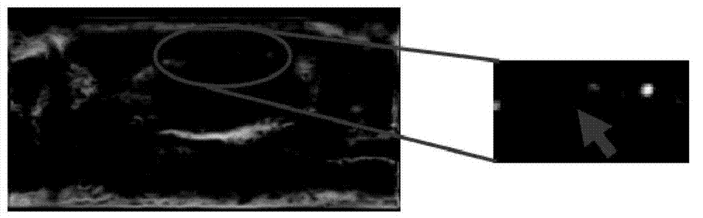
[0136] Step 2, use the formula (1) to perform edge detection on the cropped 3D image to obtain an initial edge map. In this example, a tomographic slice (12 slices) of 3D volume data is selected, such as figure 2 As shown, the right side is a partially enlarged schematic diagram of the left side image. The closer the gray value of a pixel point in the figure is...


PUM


Abstract

The invention discloses a three-dimensional mammary gland ultrasound image partition method based on homoplasmon and partial energy. The method includes that the three-dimensional mammary gland ultrasound image to be processed is inputted, the image to be processed is carried out an edge detection. A detected edge curved surface is carried out fitting. The homoplasmon and an overall energy functional are defined, and a numerical calculation is the last step. The three-dimensional mammary gland ultrasound image partition method based on homoplasmon and partial energy has the advantages that firstly, difficult problem that in the traditional method, a three dimensional neighboring window is hard to choose is solved, so that, the neighboring window is capable of varying together with the changes of spatial position of a voxel.The homoplasmon, to some extent, can guarantee that the neighborhood cannot step over an organization, can effectively differentiate the pixels with similar appearance but belonging to different organizations, and the homoplasmon is helpful for improving the extraction accuracy of the tumor outline. Secondly, defects that for a level set model based on the global information, the unevenness of the gray level is hard to overcome and a level set model based on the partial regional information is fallen into the local extremum are solved, the extraction.

Description

technical field [0001] The invention relates to the technical field of medical ultrasonic image processing, in particular to a three-dimensional mammary gland ultrasonic image segmentation method based on homogeneous body and local energy. Background technique [0002] The morbidity and mortality of breast cancer rank first among women's diseases and have become the killer of women's death. In the detection of breast tumors, ultrasonography is favored due to its advantages such as non-invasive, non-radiation, and suitable for large-scale inspections. Compared with two-dimensional breast ultrasound images, three-dimensional breast ultrasound imaging can provide more comprehensive and richer image information, which is more conducive to doctors' observation and analysis. However, there are individual differences in the reading of ultrasound images by doctors, especially for 3D images with a huge amount of data, the reading of doctors is particularly cumbersome. In order to r...

Claims


Application Information

Patent Timeline
IPC IPC(8): G06T7/00
Inventor 高梁刘晓云冯前进陈武凡
Owner UNIV OF ELECTRONIC SCI & TECH OF CHINA
Why Eureka
Try Eureka