Method and device for reducing metal artifacts in medical images

A medical image and metal artifact technology, applied in the field of image processing, can solve the problems of artifacts, inability to cover channels, and inability to ensure that large metals have smooth edges and retain small metals, etc., and achieve the effect of easy realization and good image quality

Active Publication Date: 2013-07-03
GE MEDICAL SYST GLOBAL TECH CO LLC
View PDF4 Cites 8 Cited by
  • Summary
  • Abstract
  • Description
  • Claims
  • Application Information

AI Technical Summary

Problems solved by technology

from Figure 3B and Figure 3C It can be seen that the channels with metal projections from BFP cannot cover all the real channels with metal projections
Therefore, there are residual metal artifacts in the reconstruction, such as Figure 3C shown
[0015] Third, the interpolation in the projection data correction (step 140) will result in data loss for small metals, which will introduce additional artifacts
Then, severe artifacts appear along the direction connecting small and large metals, such as Figure 4C shown
[0016] Fourth, in the metal backfill step (step 160), a single smoothing radius does not guarantee good enough image quality for both small and large metals
[0018] Therefore, the MAR method in current RT cannot guarantee that small metals are well preserved while large metals have smooth edges

Method used


Image

  • Method and device for reducing metal artifacts in medical images
  • Method and device for reducing metal artifacts in medical images
  • Method and device for reducing metal artifacts in medical images

Examples


Embodiment Construction

[0048] The present invention will be further described below in conjunction with the accompanying drawings and specific embodiments.

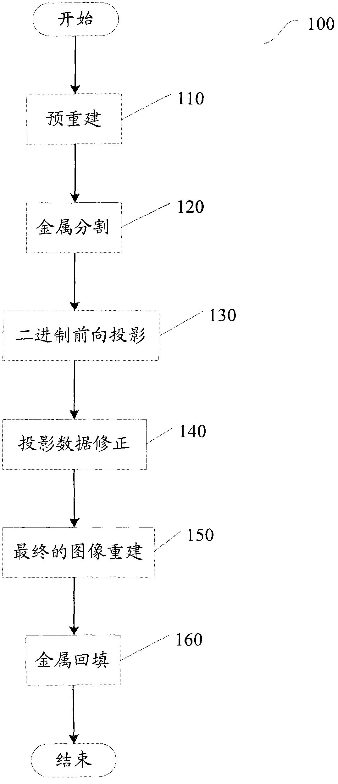
[0049] Figure 6 A flowchart of a method 600 according to the present invention is schematically shown. In method 600, artifacts generated by large metal objects and small metal objects in the same tomographic image are processed in different ways, thereby reducing metal artifacts.

[0050] Method 600 begins at step 610, where voxels belonging to small metals in the original mask are removed. This removal can be performed by erosion, or by other methods known to those skilled in the art, such as low-pass filtering. Metallic markers for tumor localization are usually smaller than 2 mm according to clinical requirements in RT. In the reconstructed image achieved with a DFOV of 50 cm, only 1–2 pixels are contained in the original metal mask obtained from threshold-based segmentation. The number of corrosions (N e ) to 1 to completely remove i...


PUM


Abstract

The invention provides a method and a device for reducing metal artifacts in medical images, wherein the same fault of medical images comprises artifacts comprises artifacts generated by large metal objects and small metal objects with different sizes. The method is used for processing artifacts generated by the large metal objects and the small metal objects in the same fault image by different methods. According to the invention, the small metal objects can be automatically excluded out of the algorithm processing, and meanwhile the artifacts caused by the large metal objects can be removed as much as possible.

Description

technical field [0001] The present invention relates generally to image processing, and more particularly to methods and apparatus for reducing metal artifacts in medical images. Background technique [0002] During radiotherapy, on the one hand, for patients with metal implants (such as dentures, alloy femoral heads), serious shadow artifacts will be produced in the obtained medical images (eg CT images). This condition is very common in patients with brain or prostate tumors. On the other hand, doctors usually use small metal markers to localize tumors for radiotherapy, which also introduces metal artifacts. In the radiotherapy of tumors, the accuracy of the tumor location provided by computed tomography (CT) images is very important, so it is necessary to reduce the influence of metal artifacts. [0003] figure 1 is a flowchart of a prior art MAR method 100 . [0004] The method starts at step 110, where a pre-reconstruction is first performed on the CT image. This p...

Claims


Application Information

Patent Timeline
Patent Type & Authority Applications(China)
IPC IPC(8): G06T5/00
Inventor 李军李硕孙智慧董加勤
Owner GE MEDICAL SYST GLOBAL TECH CO LLC
Why Eureka
Try Eureka