Ultrasonic Diagnostic Apparatus, Medical Image Processing Apparatus, And Medical Image Processing Method

A medical imaging and diagnostic device technology, applied in the directions of ultrasonic/sonic/infrasonic image/data processing, ultrasonic diagnosis, ultrasonic/sonic/infrasonic diagnosis, etc., which can solve the problems of judgment error, non-existence of blood flow, low robustness, etc.

Active Publication Date: 2015-06-17
TOSHIBA MEDICAL SYST CORP
View PDF12 Cites 0 Cited by
  • Summary
  • Abstract
  • Description
  • Claims
  • Application Information

AI Technical Summary

Problems solved by technology

However, there are no objective parameters for evaluating the morphology of blood flow
[0010] Regarding the shape of the blood flow, the above-mentioned ultrasound image is usually confirmed by an examiner who judges whether the site of interest is normal or abnormal based on experience, but this judgment may not be accurate due to time differences depending on the examiner.
In other words, the determination of whether the blood flow pattern is normal or abnormal may vary depending on the inspector, and it can be said that the robustness is low.

Method used


Image

  • Ultrasonic Diagnostic Apparatus, Medical Image Processing Apparatus, And Medical Image Processing Method
  • Ultrasonic Diagnostic Apparatus, Medical Image Processing Apparatus, And Medical Image Processing Method
  • Ultrasonic Diagnostic Apparatus, Medical Image Processing Apparatus, And Medical Image Processing Method

Examples


Embodiment Construction

[0022] Hereinafter, an ultrasonic diagnostic apparatus according to the present embodiment will be described with reference to the drawings. However, in the following description, the same reference numerals are attached to constituent elements having substantially the same function and structure, and the description will be repeated only when necessary.

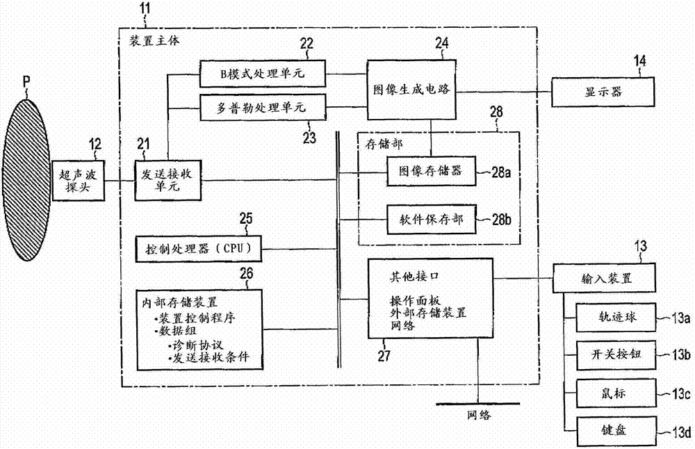
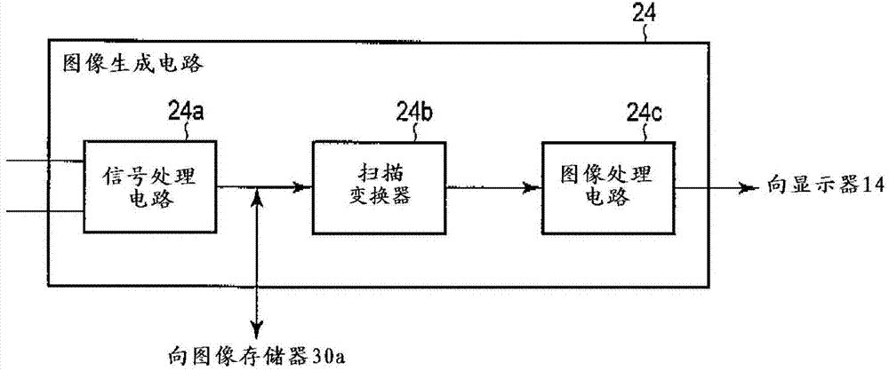
[0023] figure 1 It is a diagram showing the block configuration of the ultrasonic diagnostic apparatus 10 according to the present embodiment. Such as figure 1 As shown, the ultrasonic diagnostic apparatus 10 includes an ultrasonic diagnostic apparatus main body (hereinafter simply referred to as an apparatus main body) 11 , an ultrasonic probe 12 , an input device 13 , and a display 14 . In addition, the device main body (medical image processing device) 11 includes: a transmitting and receiving unit 21, a B-mode processing unit 22, a Doppler processing unit 23, an image generating circuit 24, a control processor (CPU) 25...


PUM


Abstract

According to one embodiment, a ultrasonic transmission / reception unit transmits / receives ultrasonic waves to / from a subject through the ultrasonic probe and generates an echo signal relating to a scan surface. An image generation unit generates an ultrasonic image relating to the scan surface based on the echo signal. A filter executes filter processing with respect to the ultrasonic image and extracts image constituent elements. A feature information generation unit generates feature information indicative of an amount of change in number of the extracted image constituent elements with respect to a change in characteristics of the filter.

Description

[0001] This application claims the priority of Japanese Patent Application No. 2012-006036 filed on January 16, 2012, and uses the entire content of the above-mentioned Japanese Patent Application in this application. technical field [0002] Embodiments of the present invention relate to an ultrasonic diagnostic apparatus, a medical image processing apparatus, and a medical image processing method. Background technique [0003] Ultrasonic diagnostic equipment is a diagnostic equipment that displays images of information in the living body. Compared with other image diagnostic equipment such as X-ray diagnostic equipment and X-ray computer tomography equipment, it is inexpensive and has no radiation. It is used for non-invasive real-time observation. Useful device to take advantage of. Ultrasonic diagnostic equipment can be used in a wide range of applications, from circulatory organs such as the heart to the abdomen such as the liver and kidneys, peripheral blood vessels, o...

Claims


Application Information

Patent Timeline
Patent Type & Authority Patents(China)
IPC IPC(8): A61B8/06A61B8/08A61B8/00
CPCA61B8/5207A61B8/14A61B8/4444A61B8/463A61B8/488A61B8/5246A61B8/5269
Inventor 姚淙神山直久川岸哲也
Owner TOSHIBA MEDICAL SYST CORP
Why Eureka
Try Eureka