Method for generating distribution image used for observing cervix tumour radiotherapy total dose

A dose distribution, uterine tumor technology, applied in the field of X-ray radiation measurement, can solve inaccurate and other problems

Inactive Publication Date: 2013-07-31
SOUTHERN MEDICAL UNIVERSITY
View PDF4 Cites 7 Cited by
  • Summary
  • Abstract
  • Description
  • Claims
  • Application Information

AI Technical Summary

Problems solved by technology

Although the above method can also obtain registration results, and the registration results can be applied to the assessment of radiotherapy radiation dose, but because this method directly uses the image containing the applicator for registration, if the obtained image is used Obviously inaccurate for radiotherapy radiation dose assessment

Method used


Image

  • Method for generating distribution image used for observing cervix tumour radiotherapy total dose
  • Method for generating distribution image used for observing cervix tumour radiotherapy total dose
  • Method for generating distribution image used for observing cervix tumour radiotherapy total dose

Examples


example 1

[0071] Example 1 (Generation of the distribution image of the total dose of radiotherapy for uterine tumors)

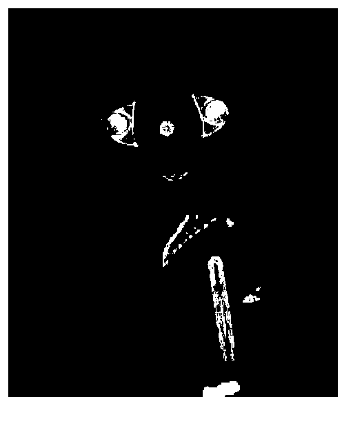
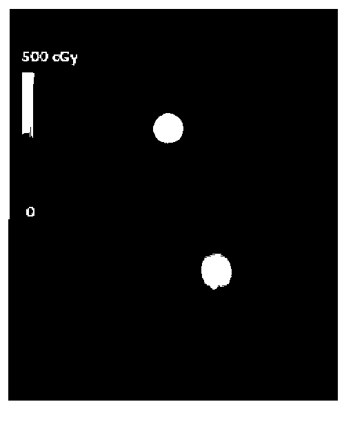
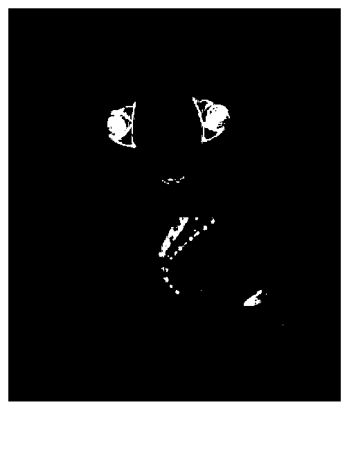
[0072] In this embodiment, the pelvic HDR CT image ( figure 1 ), pelvic HDR dose distribution image ( figure 2 ), pelvic IMRT CT images ( image 3 ) and pelvic IMRT dose distribution images ( Figure 4 ) describe the method of the present invention in detail, and its specific steps are as follows:

[0073] Step 1: Read in as figure 1 The resolutions shown are all 256*256*108 pelvic HDR CT images containing applicators and such as figure 2 The pelvic HDR dose distribution image shown and image 3 The resolutions shown are all 256*256*108 pelvic IMRT CT images without applicators and such as Figure 4 Pelvic IMRT dose distribution images shown.

[0074] Step 2: Since the applicator is metal and has a built-in source of X-ray radiation, the applicator is figure 1 The CT value of the corresponding area is greater than 200HU, that is, the pixel value is greater t...

example 2

[0091] Example 2 (analysis and comparison of effects)

[0092] The method proposed by the present invention is mainly applied to dose superposition in radiotherapy, so as to evaluate the actual total dose of radiation received by a patient during the entire radiotherapy period. In this embodiment, the dose distributions of HDR brachytherapy internal irradiation and IMRT external irradiation are shown in the attached figure 2 And attached Figure 4 shown. From attached Figure 4 It can be seen that the dose of IMRT is only the dose distribution absorbed by human tissues. Before the dose is superimposed, the IMRT dose must be expanded in the vaginal area, and then it can be superimposed on the HDR dose containing the applicator.

[0093] In order to compare the difference between the method of the present invention and existing methods, the present invention and Christensen G E (references Christensen G E, Carlson B, Chao K S, Yin P, Grigsby P W, Nguyen K, Dempsey J F, Lerma...


PUM


Abstract

The invention relates to measurement of X-ray radiation and in particular relates to a method for generating a distribution image used for observing cervix tumour radiotherapy total dose. The method comprises the following steps of: firstly dividing a source applicator region of an HDR CT (high-dose-rate brachytherapy computed tomography) image containing a source applicator to obtain a binary mask image; carrying out shrinkage distortion and interpolation on the HDR CT image by utilizing the mask image; then registering by taking the HDR CT image as a reference image and an IMRT CT (intensity modulated radiation therapy computed tomography) containing no source applicator as a floating image, and deforming the IMRT CT image and an IMRT dose distribution image by utilizing the deformation field; then amplifying a vagina region of the deformed IMRT CT image; then registering by taking the IMRT CT image as a floating image and the HDR CT image with the source applicator region to be an air CT value as a reference image; deforming the IMRT dose distribution image with the amplified vagina region by utilizing the deformation field; and finally superposing the deformation result on the a HDR dose distribution image and fusing the deformation result with the HDR CT image. By using the image generated by the method, the total dose of radiation on a patient suffering from cervix tumour can be accurately evaluated.

Description

technical field [0001] The invention relates to the measurement of X-ray radiation, in particular to a method for generating images for evaluating radiation doses of uterine tumor radiotherapy. Background technique [0002] In conventional radiotherapy, the patient's computed tomography (CT, Computed Tomography) image acquired for the first time is used as the planning image, and only the planned CT image is used to generate a treatment plan to treat the patient. But in fact, in each fractionated treatment, the patient's internal anatomical structure will change, such as the error of the technician's positioning, the peristalsis of the patient's internal organs, and the tumor will shrink with the progress of radiotherapy. Planning and implementing radiation therapy will inevitably lead to serious dose errors and may involve normal tissue into high-dose areas. Adaptive radiotherapy (ART, Adaptive Radiotherapy) technology can solve the above problems. ART technology obtains ...

Claims


Application Information

Patent Timeline
Patent Type & Authority Applications(China)
IPC IPC(8): G06T11/00A61B6/03
Inventor 甄鑫周凌宏陈海斌王琳婧胡洁肖阳
Owner SOUTHERN MEDICAL UNIVERSITY
Why Eureka
Try Eureka