
Microfluidic chip preparation method for culturing and detecting lung cancer cells

A technology of microfluidic chips and lung cancer cells, applied in biochemical equipment and methods, tissue cell/virus culture devices, specific-purpose bioreactors/fermenters, etc., can solve problems such as technologies and methods that have not been reported yet , to achieve the effects of cell attachment, cell tentacles, and volume increase

Inactive Publication Date: 2016-09-07
CHONGQING UNIV
View PDF2 Cites 0 Cited by
  • Summary
  • Abstract
  • Description
  • Claims
  • Application Information

AI Technical Summary

Problems solved by technology

However, microfluidic chip technology is still in its infancy, and its related technologies and methods for lung cancer cell culture and detection have not been reported yet.

Method used


Image

  • Microfluidic chip preparation method for culturing and detecting lung cancer cells
  • Microfluidic chip preparation method for culturing and detecting lung cancer cells
  • Microfluidic chip preparation method for culturing and detecting lung cancer cells

Examples


Embodiment Construction

[0030] Below in conjunction with accompanying drawing and example the present invention is described in further detail.

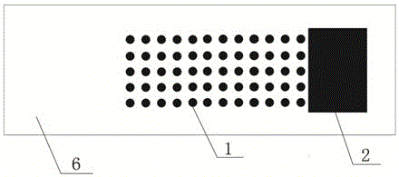
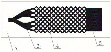
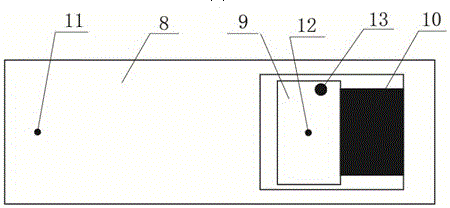
[0031] see Figure 1~Figure 8 A method for preparing a microfluidic chip for culturing and detecting lung cancer cells, comprising the steps of:

[0032] Preparation of mask: use drawing software to draw the mask diagram of cell culture layer and microchannel layer respectively, and print the mask diagram with film film to obtain the mask of cell culture layer and microchannel layer, the mask of cell culture layer and the mask of microchannel layer The shape and size of the microchannel layer mask are the same; the drawing software can use coreldraw, etc.

[0033] Among them, 60 circles are arranged in a matrix on the mask of the cell culture layer, 12 in each row, 5 rows in total, and the diameter of each circle is 1.5mm. A rectangle is arranged near one end of the matrix circle, the length of the rectangle is 16mm, and the width is 11mm. The circles an...


PUM

PropertyMeasurementUnit
sizeaaaaaaaaaa
diameteraaaaaaaaaa

Abstract

The invention discloses a method for preparing a micro-fluidic chip for culturing and detecting lung cancer cells. The method comprises the following steps: preparing a mask; preparing a hydrogel material; preparing a cell culture layer; arranging the mask of the cell culture layer on a glass substrate, and injecting the hydrogel material under shading conditions, so that the hydrogel material is cured; preparing a micro-channel layer, namely arranging the mask of the micro-channel layer on the prepared cell culture layer, and injecting the hydrogel material under the shading conditions, so that the hydrogel material is cured; and eluting and preparing a sealing cover layer, namely eluting the prepared micro-channel layer, and sealing by using PDMS. According to the micro-fluidic chip prepared by the method disclosed by the invention, multiple groups of cell samples can be simultaneously cultured, a three-dimensional growth environment is provided for the cells, the human lung tissue structure is completely simulated, a growth environment close to the human body is provided for the cells, and the metabolite is very close to the metabolite of human cells, so that the detection data is closer to the human real conditions, and early diagnosis of lung cancer is promoted.

Description

technical field [0001] The invention relates to a method for preparing a microfluidic chip, in particular to a method for preparing a microfluidic chip for cultivating and detecting lung cancer cells. Background technique [0002] Lung cancer, also known as bronchial cancer, occurs in the epithelium of the bronchial mucosa. It is one of the malignant tumors with the fastest-growing morbidity and mortality and the greatest threat to the health and life of the population. Because the symptoms of lung cancer are not obvious in the early stage, it is often discovered at an advanced stage. Therefore, early diagnosis and early treatment of lung cancer are the most effective ways to prevent and treat lung cancer and reduce the mortality rate. At present, clinically, the common diagnostic methods for lung cancer are as follows: X-ray detection, computerized tomography (CT), magnetic resonance tomography (MRI), sputum exfoliated cell examination, fiber bronchial examination, percutan...

Claims


Application Information

Patent Timeline
Patent Type & Authority Patents(China)
IPC IPC(8): C12M3/04C12M1/00
CPCC12M23/16C12M23/42C12Q1/02
Inventor 侯长军邓艳郭明遗霍丹群杨眉法焕宝罗小刚
Owner CHONGQING UNIV
Learn More