Fibrinogen compound and application thereof
A technology of fibrinogen and complexes, applied in the direction of peptide/protein components, active ingredients of hydroxyl compounds, drug combinations, etc., can solve problems such as no effect and ineffective visual function of the host
- Summary
- Abstract
- Description
- Claims
- Application Information
AI Technical Summary
Problems solved by technology
Method used
Image
Examples
Embodiment 1
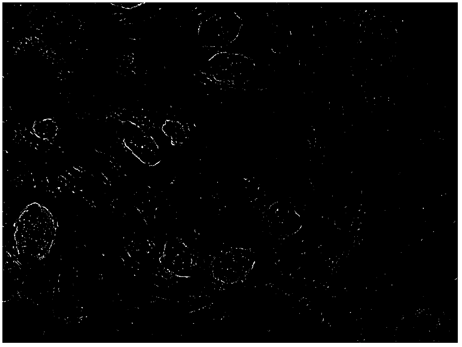
[0026] Example 1 Cell Culture and Acquisition
[0027] 1.1 About retinal ganglion cells (Retinal stem cell, RGC)
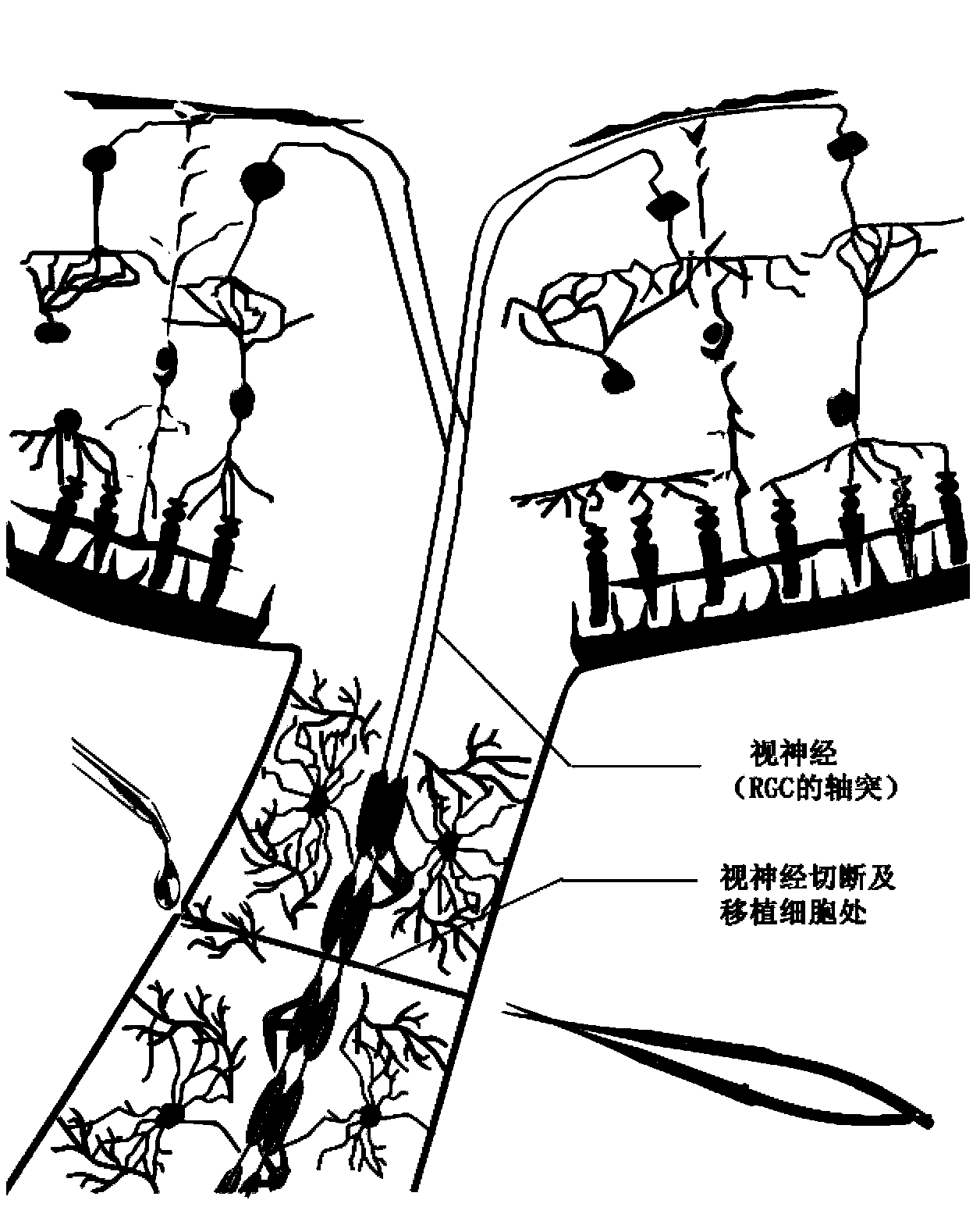
[0028] Retinal ganglion cells are located in the innermost layer of the retina, the ganglion cell layer. The axons of RGCs gather into bundles and leave the eye to form the optic nerve, which forms the optic tract with the optic nerve from the other eye at the optic chiasm, and finally projects to the lateral geniculate body. RGCs are interconnected and form a network of interactions, and their axons are also closely associated with each other. Neuronal axons absorb neurotrophic factors and transport them to the cell body through retrograde transport, so axonal necrosis can cause neuronal cell body malnutrition or even death. A very small number of RGCs or their axons degenerate into necrosis or function, which will lead to progressive dysfunction of the remaining normal RGCs and their axons, and eventually decompensation to apoptosis of all cells leading to bli...
Embodiment 2
[0054] Preparation of embodiment 2 fibrinogen complex
[0055] Add NaCl to deionized water to make 10ml of 0.8% (w / v) NaCl solution, filter through the filter head, dissolve 400mg of fibrinogen in it, add the neurotrophic factors shown in Table 1 in turn, and finally add prothrombin , to obtain the nerve regeneration biological glue of the present invention, wherein the final concentration of fibrinogen is 22-28U / ml, and the final concentration of thrombin is 22-28U / ml. The bioglue is formed about 15 minutes after preparation, has high viscosity, high mechanical strength, and large porosity, and is easier to fix at the injection site, and is suitable for injection at the site of optic nerve injury in living bodies (animals / humans).
Embodiment 3
[0056] Embodiment 3 animal experiments
[0057] 3.1 Experimental animals
[0058] Healthy Sprague Dawley rats, male or female, weighing 250g-300g, examined under a slit-lamp microscope, had double gills as large and round as L, responded well to light, had no obvious eye disease, and no crooked neck. Rats were anesthetized by intraperitoneal injection of 3% pentobarbital sodium solution, 1ml / kg, and the middle part of the upper eyelid was cut perpendicular to the eyelid margin under the binocular operating microscope until the supraorbital edge, and the bulbar conjunctiva and part of the fornix conjunctiva were cut in the same direction , expose and separate the superior rectus muscle, bluntly dissect the fascia along the direction of the superior rectus muscle to expose the optic nerve, cut the superior rectus muscle, clamp the severed end of the superior rectus muscle insertion with micro-toothed forceps, pull the eyeball downward, and fully expose the optic nerve. A specia...
PUM
 Abstract
Description
Claims
Application Information
 - R&D
- Intellectual Property
- Life Sciences
- Materials
- Tech Scout
- Unparalleled Data Quality
- Higher Quality Content
- 60% Fewer Hallucinations
Browse by: Latest US Patents, China's latest patents, Technical Efficacy Thesaurus, Application Domain, Technology Topic, Popular Technical Reports.
© 2025 PatSnap. All rights reserved.Legal|Privacy policy|Modern Slavery Act Transparency Statement|Sitemap|About US| Contact US: help@patsnap.com


