A three-dimensional visualization method of blood vessels based on intravascular ultrasound images

A technology of ultrasound imaging and ultrasound images, which is applied in the processing of 3D images, catheters, image data processing, etc., and can solve the problems of lack of image and clear local information

Inactive Publication Date: 2015-11-18
BEIJING UNIV OF TECH
View PDF3 Cites 0 Cited by
  • Summary
  • Abstract
  • Description
  • Claims
  • Application Information

AI Technical Summary

Problems solved by technology

For the 3D model obtained by volume rendering, although it can display complete 3D information, it lacks vivid and clear local information

Method used


Image

  • A three-dimensional visualization method of blood vessels based on intravascular ultrasound images
  • A three-dimensional visualization method of blood vessels based on intravascular ultrasound images
  • A three-dimensional visualization method of blood vessels based on intravascular ultrasound images

Examples


Embodiment Construction

[0050] The present invention is realized by adopting the following technical means:

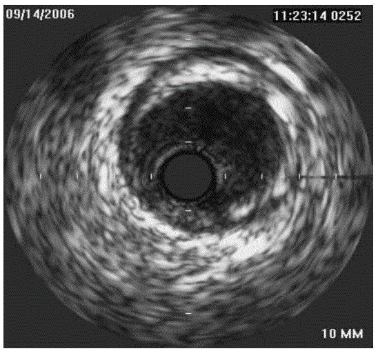
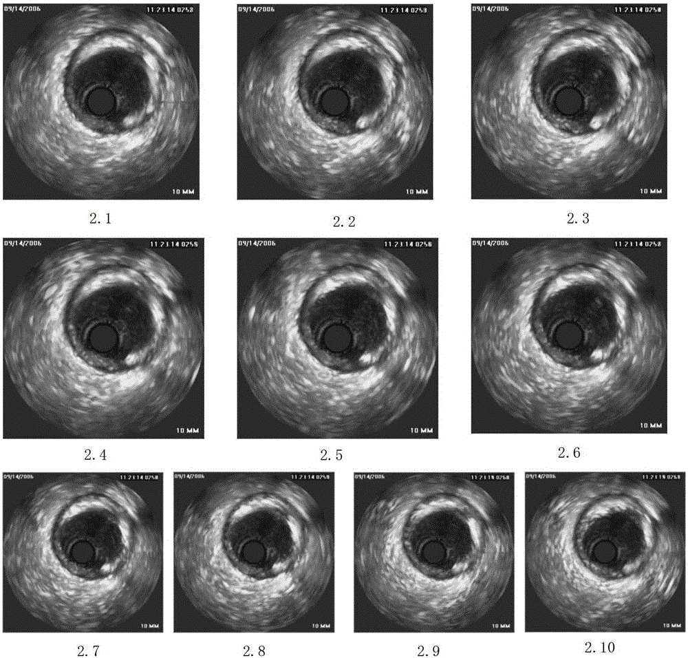
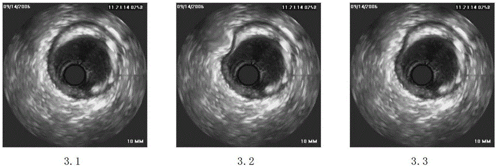
[0051] A three-dimensional visualization method of blood vessels based on intravascular ultrasound images. Firstly, the intravascular ultrasound image sequence is denoised by combining image sequence averaging, median filtering and wavelet soft threshold denoising methods, and then the quadratic polynomial is used to fit the image deformation to realize ultrasound image registration and compensate the image sequence acquisition process Finally, the reconstruction of the three-dimensional model of the blood vessel and the plane section at any angle are realized by using the ray-casting algorithm and the slice reorganization method, so as to obtain an intuitive and vivid three-dimensional visualization effect of the blood vessel.

[0052] The above three-dimensional visualization method of blood vessels based on intravascular ultrasound images comprises the following steps:

[0053] Step 1. Us...


PUM


Abstract

The invention provides a vessel three-dimensional visualized method based on an ultrasonic image in a vessel, and relates to the field of computer medical image analysis. The method is characterized in that firstly, noise reduction is carried out on an image sequence through combination of the multi-image average de-noising method, the median filtering method and the wavelet soft-threshold de-noising method, the vessel three-dimensional visualized method can lower image noise and well maintain important detail information of the image, and noise reduction efficiency of the image is high; secondly, image registration is achieved due to the fact that a quadratic polynomial is used for image deformation fitting so as to compensate deformation generated during collection of the image sequence; thirdly, a three-dimensional vessel model is drawn by means of the ray-casting algorithm; finally, plane cutting of the three-dimensional vessel model at any angle is achieved by means of a slicing reassembling method, internal structure information of the vessel is displayed, and conditions are provided for lesion analysis.

Description

technical field [0001] The invention relates to the field of computer medical image analysis, in particular to a blood vessel three-dimensional visualization method based on intravascular ultrasonic images. Background technique [0002] Intravascular ultrasound image is a kind of tomographic slice image, which can display the cross-sectional view of the blood vessel at the current position, describe the tissue composition of the blood vessel wall, lumen and plaque in detail, and then calculate the lumen diameter and cross-sectional area of ​​the blood vessel, and can be based on The histological analysis of the plaque acoustic characteristics revealed early atherosclerotic plaques, which have an incomparable advantage in the diagnosis of plaque lesions compared with coronary angiography. In clinical practice, for the Intravascular Ultrasound (IVUS) image sequence of a certain segment of blood vessels, doctors are not limited to observing each frame of images individually, bu...

Claims


Application Information

Patent Timeline
Patent Type & Authority Patents(China)
IPC IPC(8): G06T17/00G06T15/00A61B8/12
Inventor 汪友生吴焕焕苗晓静王志东董路李冠宇
Owner BEIJING UNIV OF TECH
Why Eureka
Try Eureka