Method for calculating, displaying and storing radiation dosages of CT image formation

A radiation dose, CT imaging technology, applied in dosimeters, computed tomography scanners, echo tomography, etc., can solve the problem of not being able to count the historical cumulative radiation dose of X-ray radiation dose of a single tissue or organ

Inactive Publication Date: 2013-10-09
奚岩
View PDF10 Cites 26 Cited by
  • Summary
  • Abstract
  • Description
  • Claims
  • Application Information

AI Technical Summary

Problems solved by technology

The patent application discloses a CT imaging radiation dose display system, but the content of the patent application is limited to displaying the total radiation dose after the CT scan, and can

Method used


Image

  • Method for calculating, displaying and storing radiation dosages of CT image formation
  • Method for calculating, displaying and storing radiation dosages of CT image formation
  • Method for calculating, displaying and storing radiation dosages of CT image formation

Examples


Embodiment 1

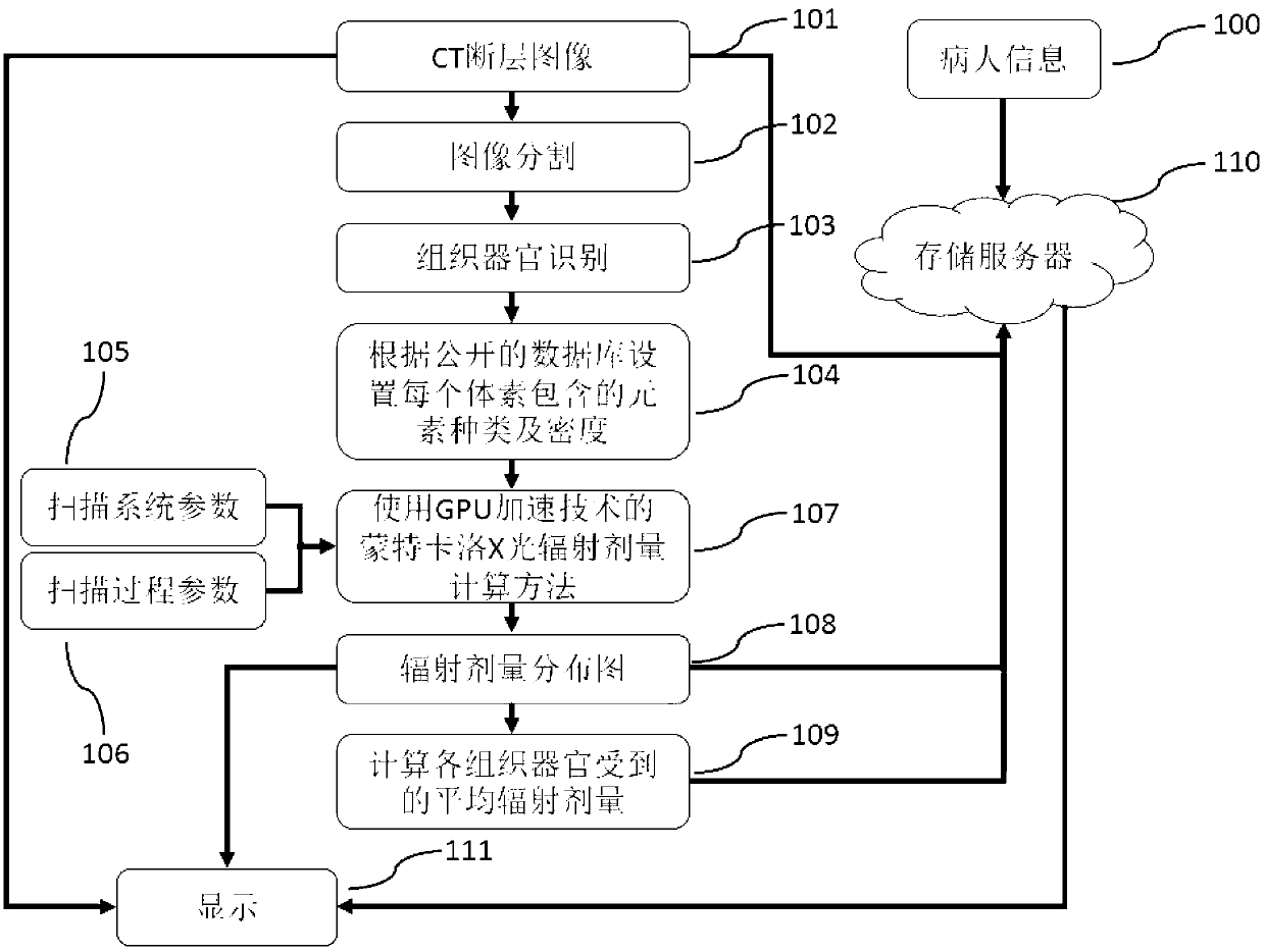
[0033] Embodiment 1: Perform an abdominal CT scan on patient A, and then calculate, display, and store the radiation dose of this CT scan.

[0034] see figure 1 , according to the implementation process of the inventive method comprising:

[0035] 1. Obtain the system parameters of patient A for CT scanning ( figure 1 in step 105) and scanning process parameters ( figure 1 in step 106).

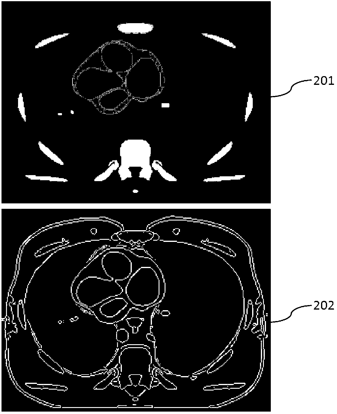
[0036] 2. According to the input CT image ( figure 1 In step 101), information such as CT value information and structural shape are used to perform three-dimensional image segmentation on the CT image ( figure 1 Step 102 in step 102), and identify the tissue and organ category corresponding to the segmented region ( figure 1 in step 103). Such as figure 2 As shown, 201 in the figure is a tomographic image obtained by CT scanning, and 202 is a representation diagram of tissues and organs after the tomographic image is segmented and marked.

[0037] 3. Correspond the classified ti...

Embodiment 2

[0041] Embodiment 2: Before performing abdominal CT scan on patient B, adjust the parameters of this CT scan appropriately according to the historical records of X-ray CT radiation dose received by patient B.

[0042] see figure 1 , according to the implementation process of the inventive method comprising:

[0043] 1. The doctor who performs CT scan imaging on patient B first sends the personal information of patient B and the CT scan information ( figure 1 In step 100) input to the storage server of CT radiation dose ( figure 1 in step 110).

[0044] 2. According to the record of patient B on the storage server, the X-ray CT radiation record received by the patient is displayed on the doctor's operation interface, such as Figure 4 shown. It includes: the basic information of the patient ( Figure 4 401); all the CT scans of the patient and the CT scans in the past year ( Figure 4 middle 402); the patient’s cumulative radiation dose to the tissues and organs that m...


PUM


Abstract

The invention discloses a method for calculating, displaying and storing radiation dosages of CT image formation. The method comprises the steps of, in CT scanning, calculating the average radiation dosage received by each tissue and each organ in the CT scanning, calculating the distribution of obtained radiation dosages, and uploading relevant information to a storage server; simultaneously displaying historical accumulated radiation dosages of relevant tissues and organs of the scanning according to all radiation dosage data, stored in the storage server, of a patient. The method uses tissues and organs as units, uses historical data, can monitor accumulated radiation dosages received by a specific organ of the patient, reminds the patient of targeted body examination after a threshold is surpassed, and reminds a doctor of proper adjustment of CT scanning parameters, so that the purpose of controlling accumulated radiation dosages of tissues and organs is further achieved.

Description

technical field [0001] The invention belongs to the technical field of CT imaging, and relates to methods for calculating, displaying and storing radiation doses of CT imaging. Background technique [0002] CT imaging technology is a method to invert the internal structure of the object by measuring the attenuation signal of the scanned object to the incident X-ray at multiple angles. A CT imaging device for medical diagnosis mainly includes the following parts: an X-ray tube, an X-ray detector and a frame for placing the X-ray tube and the detector. During a CT scan, the gantry rotates around the patient while a computer program controls the emission of X-rays and the acquisition of data by the detectors. CT imaging technology is based on the principle of attenuation of incident X-rays by human tissue. X-rays have certain radiation damage to the human body. If the human body receives too much X-ray exposure, it may even cause cancer. With the widespread use of CT imaging ...

Claims


Application Information

Patent Timeline
IPC IPC(8): A61B6/03G01T1/02
Inventor 奚岩
Owner 奚岩
Why Eureka
Try Eureka