
Maximum a posteriori reconstruction method for pet images based on joint prior model of anatomical function

A combined prior and maximum a posteriori technique, applied in the field of PET image processing of medical images, can solve problems such as affecting the quality of reconstructed images, large noise errors in segmentation or edge extraction, etc.

Active Publication Date: 2017-05-03
广东高尚医学影像诊断中心有限公司
View PDF2 Cites 0 Cited by
  • Summary
  • Abstract
  • Description
  • Claims
  • Application Information

AI Technical Summary

Problems solved by technology

[0005] However, most of the existing anatomical prior-guided maximum a posteriori reconstruction techniques are based on the edge or region information of the anatomical image. First, the anatomical image needs to be segmented or edge extracted. However, there is no absolutely robust method for anatomical image segmentation or edge extraction. , there is a large noise error in segmentation or edge extraction, which seriously affects the quality of the reconstructed image

Method used


Image

  • Maximum a posteriori reconstruction method for pet images based on joint prior model of anatomical function
  • Maximum a posteriori reconstruction method for pet images based on joint prior model of anatomical function
  • Maximum a posteriori reconstruction method for pet images based on joint prior model of anatomical function

Examples


Embodiment 1

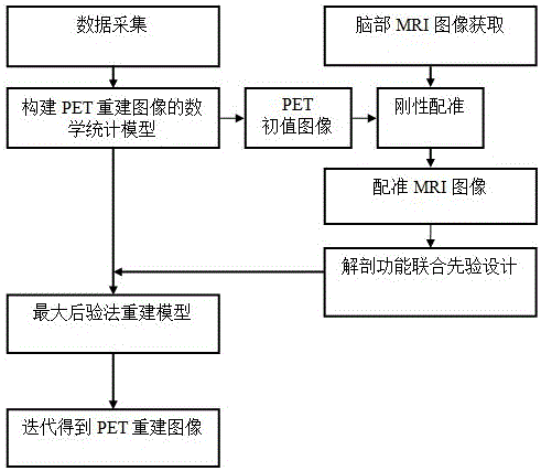
[0052] A method for maximum posterior reconstruction of PET images based on anatomical function combined prior models includes the following steps in sequence.

[0053] (1) Acquire PET data for reconstruction through imaging equipment.

[0054] Specifically, the detection data before PET imaging is collected by the imaging device, and the correction parameter values ​​and the system matrix of the imaging device are obtained at the same time, and the acquired detection data is corrected by the imaging device to obtain the corrected detection data, and the corrected detection data is obtained. The detection data is used as PET data for reconstruction. In the art, the detection data is also called projection data, and the corrected detection data or projection data is the PET data used for reconstruction.

[0055] (2) According to the statistical characteristics of the PET data obtained in step (1), construct a mathematical statistical model for reconstructing the image. PET data Mee...

Embodiment 2

[0084] Taking the brain image of the phantom shown in Fig. 2 as an example, a method for maximum posterior reconstruction of a PET image based on an anatomical function combined prior model of the present invention will be described. figure 1 As shown, the following steps are included.

[0085] (1) Acquire PET data for reconstruction through imaging equipment.

[0086] The detection data before PET imaging is collected by the imaging device, and various data correction parameter values ​​in the imaging device and the system matrix of the imaging device are obtained at the same time. In this embodiment, the data acquisition method is full three-dimensional acquisition; the system matrix P corresponds to the parallel belt-shaped geometric model. Specifically, the collected data is stored in the array first, and the system obtains the scan time calibration coefficient, the detector efficiency, the attenuation coefficient and the time correction coefficient, and all the detected random...


PUM


Abstract

Disclosed is a PET image maximum posterior reconstruction method based on a united prior model with a dissection function. The method comprises the steps of (1) obtaining reconstructed PED data; (2) constructing a mathematical statistics model used for image reconstruction; (3) solving the mathematical statistics model obtained in the step (2) by means of maximum likelihood-expectation maximization to obtain a PET initial value image; (4) carrying out registration on a pre-acquired MRI image and the PET initial value image obtained in the step (3); (5) reconstructing the mathematical statistics model, constructed in the step (2), of the PET image by means of the maximum posterior method according to the registration MRI image and PET initial value image united prior model obtained in the step (4) to obtain an optimization equation with a constraint objective function; (6) carrying out iterative computation on the optimization equation with the constraint objective function to obtain a reconstructed PET image. According to the PET image maximum posterior reconstruction method based on the united prior model with the dissection function, noise generated during PET image reconstruction can be restrained, and reconstructed image quality can be improved.

Description

Technical field [0001] The invention relates to the technical field of PET image processing of medical images, in particular to a PET image maximum posterior reconstruction method based on an anatomical function joint prior model. Background technique [0002] Positron emission tomography (PET), as a non-interventional tool for quantitatively studying the functional activity of living organisms, is becoming more and more widely used in clinical diagnosis, especially early diagnosis of diseases. [0003] However, because the collected data during PET scanning is affected by noise and other physical factors, PET image reconstruction is a pathological problem. Statistical image reconstruction methods, such as maximum likelihood-expectation maximization (ML-EM), can better consider the physical effects of the system model and can establish mathematical models for the statistical characteristics of detection data and noise. The quality of the reconstructed image is better than the trad...

Claims


Application Information

Patent Timeline
Patent Type & Authority Patents(China)
IPC IPC(8): G06T11/00
Inventor 路利军马建华胡德斌冯前进陈武凡
Owner 广东高尚医学影像诊断中心有限公司
Learn More