Eureka AIR delivers breakthrough ideas for toughest innovation challenges, trusted by R&D personnel around the world.

Extraction method of tumor elasticity features based on ultrasound elastography

A technology of elastic features and extraction methods, applied in the field of image processing, can solve the problems of large subjective influence of the diagnosers, lack of objectivity and scientificity, and inability to expand, so as to achieve accurate and scientific diagnosis and eliminate subjective differences.

Active Publication Date: 2016-12-21
SHENZHEN INST OF ADVANCED TECH CHINESE ACAD OF SCI
View PDF2 Cites 0 Cited by
  • Summary
  • Abstract
  • Description
  • Claims
  • Application Information

AI Technical Summary

Problems solved by technology

[0003] I am interested in the elastic characteristic parameters based on ultrasonic transient elastography technology proposed by "Medical Liberation Army Journal", Vol. 36, No. 11, pp. 1131-1133, 2011, and "Chinese Medical Imaging Technology", Vol. 28, No. 3, No. 3, pp. 529-533, 2012. However, the instantaneous elasticity detection technology used is a one-dimensional imaging technology. Although it can give quantitative average elastic modulus values ​​of tissues, it cannot be extended to two-dimensional elastography to obtain tissue elastic distribution information. Generally, only Suitable for detection of diffuse lesions; "Chinese Medical Imaging Technology", Volume 26, Issue 9, 2010, pages 1682-1684, "Modern Biomedicine Progress", Volume 26, Issue 9, 2010, pages 492-494, and "West China Medicine" 2010 Volume 25, No. 2, pp. 294-297, proposed the elasticity image distribution characteristics based on ultrasound real-time elastography technology. Although the most widely used elastic features in clinical diagnosis are given, real-time elastography technology is a quasi-static Compression elastography can only give relative displacement / strain diagrams, but cannot give specific values ​​of tissue local hardness, which lacks objectivity and scientificity; "Chinese Medical Imaging Technology", Volume 18, Issue 7, 2009, 589-591 The elastic characteristic strain rate ratio based on ultrasonic real-time elastography technology proposed by Page and "World Chinese Journal of Digestion" 2010, Vol. 18, No. 30, pp. Extracted from the displacement / strain map, although it can more objectively and quantitatively reflect the degree of hardness change of the lesion than the simple elastic distribution feature, but this feature is also unable to make a horizontal comparison between different lesions, such as the malignant liver mass on the basis of liver cirrhosis There is a certain overlap between the strain rate ratio intervals obtained for benign space-occupying lesions and benign liver lesions, and it is easy to cause misdiagnosis by only relying on the strain rate ratio
[0004] Therefore, the traditional ultrasound elastography technology has many limitations, which make the acquired elastic characteristics qualitative or semi-quantitative, lack of objectivity and repeatability, cannot be quantitatively analyzed, and are subject to the subjective influence of the diagnostician, so it is impossible to establish a recognized and standardized method. diagnostic criteria, which limits the wide application of ultrasound elastography in clinical practice

Method used


Image

  • Extraction method of tumor elasticity features based on ultrasound elastography
  • Extraction method of tumor elasticity features based on ultrasound elastography
  • Extraction method of tumor elasticity features based on ultrasound elastography

Examples


Embodiment Construction

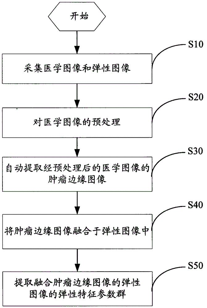
[0023] see figure 1 , a method for extracting tumor elasticity features of elastography, the specific steps are as follows:

[0024] Step S10: Input medical images and elastic images.

[0025] Elastic imaging technology is a relatively mature imaging technology, according to which elastic images can be obtained, and medical images can be B-ultrasound images or CT images or MRI (magnetic resonance) images or X-ray (X-ray) images, in the present invention In the provided embodiments, the medical image is preferably a B-ultrasound image.

[0026] Step S20: preprocessing the medical image.

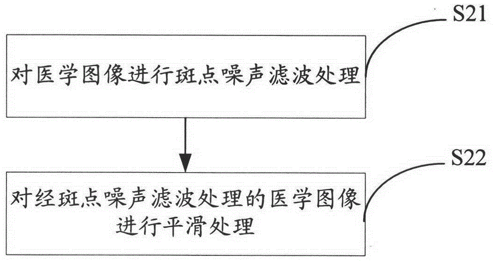
[0027] see figure 2 , the flow chart of B-ultrasound image preprocessing provided by the embodiment of the present invention, step S20 is specifically:

[0028] Step S21: Perform speckle noise filtering processing on the medical image.

[0029] The coherent nature of ultrasound imaging leads to inherent speckle noise in B-ultrasound images. Speckle noise reduces the image quality, especi...


PUM


Abstract

The invention provides a method for extracting tumor elasticity features based on ultrasound elastography, comprising the following steps: collecting medical images and elastic images; preprocessing the medical images; automatically extracting the tumor from the preprocessed medical images edge information; fusing the tumor edge information into the elastic image; extracting an elastic characteristic parameter group of the elastic image fused with the tumor edge information. The above method for extracting tumor elastic features based on elastography uses medical images to extract tumor edge information, and at the same time fuses the tumor edge information into the elastic image, and creates and quantifies the elastic feature parameter group based on the elastic image. The elastic feature parameter group can Describe the properties of the tumor from different angles, thereby eliminating the subjective differences of clinicians and making the diagnosis more accurate and scientific.

Description

technical field [0001] The invention relates to image processing technology, in particular to a method for extracting tumor elasticity features based on ultrasound elastography. Background technique [0002] Elastography is to apply an external stimulus to the tissue. Under the action of physical laws such as pressure-deformation, the tissue will produce a response, and an elastography map will be obtained according to the response. Since the concept of elastography was proposed, ultrasound elastography technology has developed rapidly in the past ten years. [0003] I am interested in the elastic characteristic parameters based on ultrasonic transient elastography technology proposed by "Medical Liberation Army Journal", Vol. 36, No. 11, pp. 1131-1133, 2011, and "Chinese Medical Imaging Technology", Vol. 28, No. 3, No. 3, pp. 529-533, 2012. However, the instantaneous elasticity detection technology used is a one-dimensional imaging technology. Although it can give quantita...

Claims


Application Information

Patent Timeline
Patent Type & Authority Patents(China)
IPC IPC(8): G06T7/00G06T5/00
Inventor 肖杨郑海荣钱明王丛知
Owner SHENZHEN INST OF ADVANCED TECH CHINESE ACAD OF SCI
Why Eureka
Eureka Blog
Learn More