Magnetoencephalography source imaging

A magnetoencephalography and imaging technology, applied in the field of imaging technology, can solve problems such as inability to diagnose neuron damage

Inactive Publication Date: 2014-04-09
RGT UNIV OF CALIFORNIA
View PDF3 Cites 26 Cited by
  • Summary
  • Abstract
  • Description
  • Claims
  • Application Information

AI Technical Summary

Problems solved by technology

Although some techniques such as diffusion tensor imaging (DTI) have shown superior sensitivity to X-ray, CT, and MRI in detecting neuronal damage (e.g., D...

Method used


Image

  • Magnetoencephalography source imaging
  • Magnetoencephalography source imaging
  • Magnetoencephalography source imaging

Examples


Embodiment Construction

[0023] Magnetoencephalography (MEG) is a technique used to map brain activity by recording the magnetic fields generated by electrical currents within cells in the brain. For example, synchronous electrical currents generated by neuronal cells can generate magnetic fields. For example, these magnetic signals can arise from the net effect of ionic currents flowing between neurons (e.g., during synaptic conduction), which can be modeled as electric dipoles, e.g., with position, orientation and amplitude currents and other non-dipole current sources that generate magnetic signals. Magnetic fields generated by neurons exhibit amplitudes on the order of femto-Tesla (fT), e.g., 10 for cortical activity. 1 fT and 10 for human alpha rhythm 3 fT. These neuronal magnetic signals can be detected by sensitive magnetometers. However, these neuronal magnetic signals are relatively weak compared to the typical ambient magnetic noise of the external body environment (which can be, for exa...


PUM


Abstract

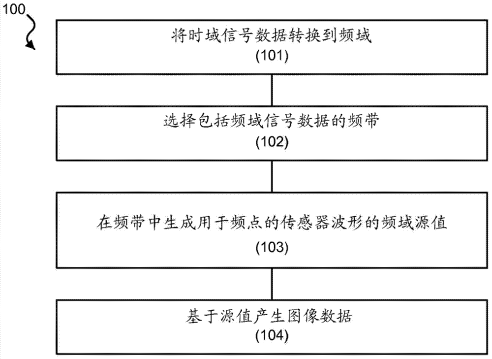
Techniques, devices and systems are disclosed for magnetoencephalography (MEG) source imaging. In one aspect, a method includes selecting signal data associated with one or more frequency bands from a spectrum of the signal data in the frequency domain, in which the signal data represents magnetic signals emitted by a brain of a subject and detected by a plurality of sensors outside the brain, defining locations of sources within the brain that generate the magnetic signals, in which the number of locations of the sources is selected to be greater than the number of sensors, and generating a source value of signal power based on the selected signal data corresponding to a respective location of the locations at the one or more frequencies.

Description

[0001] priority claim [0002] This patent document claims priority to US Provisional Application No. 61 / 489,667, entitled "MAGNETOENCEPHALOGRAPHY SOURCE IMAGING," filed May 24, 2011, the entire contents of which are incorporated herein by reference. [0003] Statement Regarding Federally Sponsored Research or Development [0004] This invention was made with government support under grants NEUC-044-06S and NURC-022-10F awarded by the US Department of Veterans Affairs. The government has certain rights in this invention. technical field [0005] This patent document relates to imaging technology. Background technique [0006] Axonal damage is a major factor in neuronal damage such as mild traumatic brain injury (TBI), early multiple sclerosis (MS), early Alzheimer's disease / dementia ( AD). Furthermore, abnormal functional connectivity exists in these neuronal disorders as well as in others such as post-traumatic stress disorder (PTSD). Neuroimaging tools have been used ...

Claims


Application Information

Patent Timeline
IPC IPC(8): A61B5/0476A61B5/055G06F19/00
CPCG01R33/4806A61B5/04009G06F19/321G01R33/56341G01R33/5602A61B5/055A61B5/7257G01R33/4808A61B5/246G16H30/40G16H50/20A61B5/4076A61B5/245
Inventor 黄明雄罗兰·R·李
Owner RGT UNIV OF CALIFORNIA
Why Eureka
Try Eureka