Application of fluorescent in-situ hybridization polyclonal separating probe for renal carcinoma and kit thereof

A technique of fluorescence in situ hybridization and polyclonal separation, which is applied in the application field of fluorescence in situ hybridization probes, can solve problems such as being unsuitable for wide application and complex technology, and achieve the effects of optimizing diagnostic methods, strong fluorescent signals, and expanding scope.

Active Publication Date: 2014-07-30
THE AFFILIATED DRUM TOWER HOSPITAL MEDICAL SCHOOL OF NANJING UNIV
View PDF3 Cites 11 Cited by
  • Summary
  • Abstract
  • Description
  • Claims
  • Application Information

AI Technical Summary

Problems solved by technology

However, in clinical work, it is impossible to routinely culture tumor cells to the mitotic stage before performing karyotype analysis, and the extraction of RNA from paraffin sections by RT-PCR is restricted by many factors and the technology is complicated and not suitable for wide application. Therefore, it is urgently needed A method for rapid, economical and accurate diagnosis of Xp11.2 translocation / TFE3 gene fusion-related renal cell carcinoma

Method used


Image

  • Application of fluorescent in-situ hybridization polyclonal separating probe for renal carcinoma and kit thereof
  • Application of fluorescent in-situ hybridization polyclonal separating probe for renal carcinoma and kit thereof
  • Application of fluorescent in-situ hybridization polyclonal separating probe for renal carcinoma and kit thereof

Examples


Embodiment 1

[0043] Step 1: Preparation of polyclonal DNA probes:

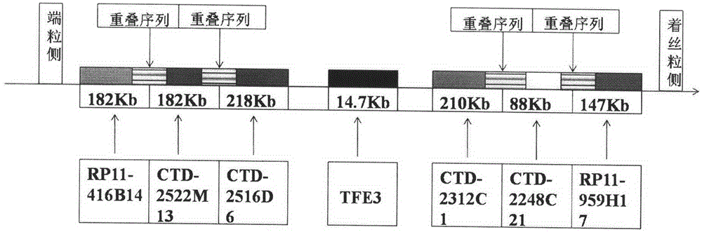
[0044] Use http: / / genome.ucsc.edu / to search for bacterial artificial chromosomes (BAC clones) corresponding to both sides of the TFE3 gene on the X chromosome (ie, the telomeric side and the centromere side), and select 3 BAC clones of similar size on both sides Fragments, control the farthest distance between the BAC clone fragments on both sides within 1500Kb and do not overlap each other, while the three fragments on the same side have a certain sequence overlap with each other. According to the above requirements, the BAC clone fragments on the telomeric side of TFE3 were selected as CTD-2516D6 (chrX: 48265836-48484403, the fragment length is about 218Kb), CTD-2522M13 (chrX: 48420425-48602847, the fragment length is about 182Kb), RP11-416B14 ( chrX: 48580872-48763198, the fragment length is about 182Kb); TFE3 centromeric BAC clone fragments are CTD-2312C1 (chrX: 48959513-49170367, the fragment length is about 210Kb), ...

Embodiment 2

[0056] Example 2: Xp11.2 Translocation / TFE3 Gene Fusion-Related Kidney Cancer Diagnostic Kit

[0057] Fluorescence in situ hybridization polyclonal separation probe kit for kidney cancer, the kit is composed of probe hybridization liquid and 4', 6-diamidino-2-phenylindole counterstain, characterized in that:

[0058] (1) TFE3 telomeric side BAC clone fragments are a combination of RP11-416B14, CTD-2522M13 and CTD-2516D6, labeled with green fluorescence; TFE3 centromere side BAC clone fragments are CTD-2312C1, CTD-2248C21 and RP11-959H17 combination, labeled red fluorescence.

[0059] (2) The two kinds of polyclonal separation probes were mixed with Human Cot-1 DNA, hybridization buffer, and purified water in proportion to prepare a probe hybridization solution, and stored in a freezer at -20°C in the dark.

[0060] (3) 4′,6-diamidino-2-phenylindole counterstain is mainly used for nuclear DNA staining.

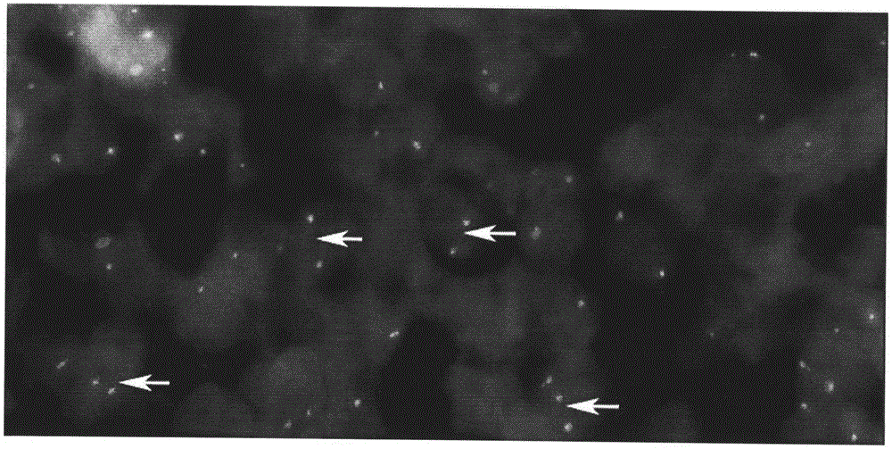
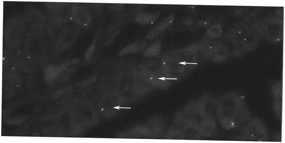
[0061] The fluorescent in situ hybridization polyclonal separation probe...


PUM


Abstract

The invention relates to application of a fluorescent in-situ hybridization polyclonal separating probe for renal carcinoma and a kit thereof. Clonal fragments used by the polyclonal separating probe provided by the invention are respectively a combination of RP11-416B14, CTD-2522M13 and CTD-2516D6, and a combination of CTD-2312C1, CTD-2248C21 and RP11-959H17. According to the invention, the defect that karyotype analysis can not be performed conventionally after tumor cells are subjected to cell culture until a mitotic phase in the prior art is overcome. The typical gene modification in Xp11.2 translocation/TFE3 gene fusion related renal carcinoma is detected by using the FISH technology, thereby diagnosing the tumor. The fluorescent in-situ hybridization polyclonal separating probe application is high in accuracy, high in specificity, high in success rate, strong in fluorescent signals and convenient to operate. The invention can be applied in paraffin sections, widens the scope of detection specimens, and greatly optimizes the diagnosis method of Xp11.2 translocation/TFE3 gene fusion related renal carcinoma.

Description

technical field [0001] The invention belongs to the field of application of fluorescent in situ hybridization probes, in particular to the application of fluorescent in situ hybridization polyclonal separation probes for kidney cancer and a kit thereof. Background technique [0002] The so-called kidney cancer refers to Xp11.2 translocation / TFE3 gene fusion-related kidney cancer, which is a subtype of kidney cancer newly named by WHO in 2004. The TFE3 gene breaks and translocates at the XP11.2 locus, and is associated with APSL and PRCC. , PSF, NonO, CLTC and other related genes are characterized by fusion, causing abnormal expression of related regulatory proteins mainly TFE3 protein. Xp11.2 translocation / TFE3 gene fusion-related RCC mainly occurs in adolescents, accounting for about 1 / 3 of RCC in children, and it is rare in adult patients. Due to its low incidence, the current understanding of Xp11.2 translocation / TFE3 gene fusion-related renal cell carcinoma is not suffi...

Claims


Application Information

Patent Timeline
Patent Type & Authority Applications(China)
IPC IPC(8): C12Q1/68C12N15/11
CPCC12Q1/6841C12Q2543/10C12Q2563/107C12Q2563/173
Inventor 甘卫东陈显成叶庆陈绍宇郭宏骞李冬梅
Owner THE AFFILIATED DRUM TOWER HOSPITAL MEDICAL SCHOOL OF NANJING UNIV
Why Eureka
Try Eureka