Mode body used for PET imaging system detector normalization correction

An imaging system and detector technology, applied in the field of medical imaging, can solve problems such as increasing the difficulty of manufacturing, increasing the complexity of the preservation process, and increasing the complexity of mechanical design, and achieves the effect of simplifying mechanical design requirements.

Inactive Publication Date: 2015-01-21
JIANGSU SINOGRAM MEDICAL TECH CO LTD
View PDF2 Cites 3 Cited by
  • Summary
  • Abstract
  • Description
  • Claims
  • Application Information

AI Technical Summary

Problems solved by technology

[0005] In order to ensure the correctness of the normalization correction before leaving the factory, it is necessary to ensure that all detection units in the PET system can be uniformly irradiated for a long time in the test, which brings great difficulties to the processing and mechanical design of the rod source:
[0006] 1. The radioactivity of the solid rod source needs to be very uniform, which makes the production cost very high
[0007] 2. In order to cover the axial field of view of the detector, the length of the rod source usually needs to exceed 20cm, which further increases the difficulty of fabrication
[0008] 3

Method used


Image

  • Mode body used for PET imaging system detector normalization correction

Examples


Embodiment Construction

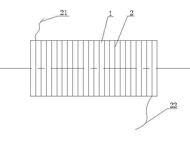
[0025] Such as figure 1 Shown, the present invention is provided with a cylinder 1, offers helical groove equidistantly on the outer surface of cylinder 1, is provided with the conduit 2 equidistantly outside cylinder 1 in spiral groove, the two ends 21 of conduit 2 and 22 are respectively provided with open ends.

[0026] In use, both ends of the conduit 2 are connected to a programmable pressure-controlled air pump. The entire cylinder 1 is fixed on the support, and laser positioning is used to precisely align the geometric axis of the cylinder with the PET detector ring.

[0027] The diameter of the cylinder 1 needs to be large enough to cover the cross-sectional field of view of the PET detector, and the length needs to be greater than the axial field of view of the PET detector.

[0028] The materials of cylinder 1 and conduit 2 should be carefully selected to ensure that they have a very low reaction cross-section within the effective energy window for concurrent event...


PUM


Abstract

The invention relates to a mode body used for PET imaging system detector normalization correction, and relates to the technical field of medical imaging. The mode body comprises a cylinder, the cylinder and a PET detector are coaxial in the axial direction, spiral grooves are formed in the outer surface of the cylinder in an equal-distance mode, guide pipes which are wound outside the cylinder in an equal-distance mode are arranged in each spiral groove, and opening ends are arranged at the two ends of each guide pipe respectively. According to the mode body, on one hand, an even stick source which is expensive does not need to be customized; on the other hand, the mechanical design requirement is simplified, and the requirement that all detection units are evenly irradiated in normalization correction is easily met.

Description

technical field [0001] The invention relates to the technical field of medical imaging, in particular to a phantom design that can be used for detector normalization correction of a positron emission computed tomography imaging system. Background technique [0002] In the field of nuclear medicine, PET (Positron Emission Tomography) is an inspection imaging technique that can non-invasively display the function and metabolism of human organs. In clinical practice, this technique can be used to guide cancer treatment. The principle is: the malignant tumor tissue in the patient's body has a strong metabolism, and the metabolites (such as glucose, protein, etc.) accumulate more. We can use radionuclides (such as 18 F. 11 C, etc.) Mark the metabolites, and then use the PET imaging system to accurately display the aggregation and activity of metabolites through three-dimensional imaging technology to reflect the activities of life metabolism. Thus, PET provides a direct and ti...

Claims


Application Information

Patent Timeline
IPC IPC(8): A61B6/03
Inventor 李楠
Owner JIANGSU SINOGRAM MEDICAL TECH CO LTD
Why Eureka
Try Eureka