

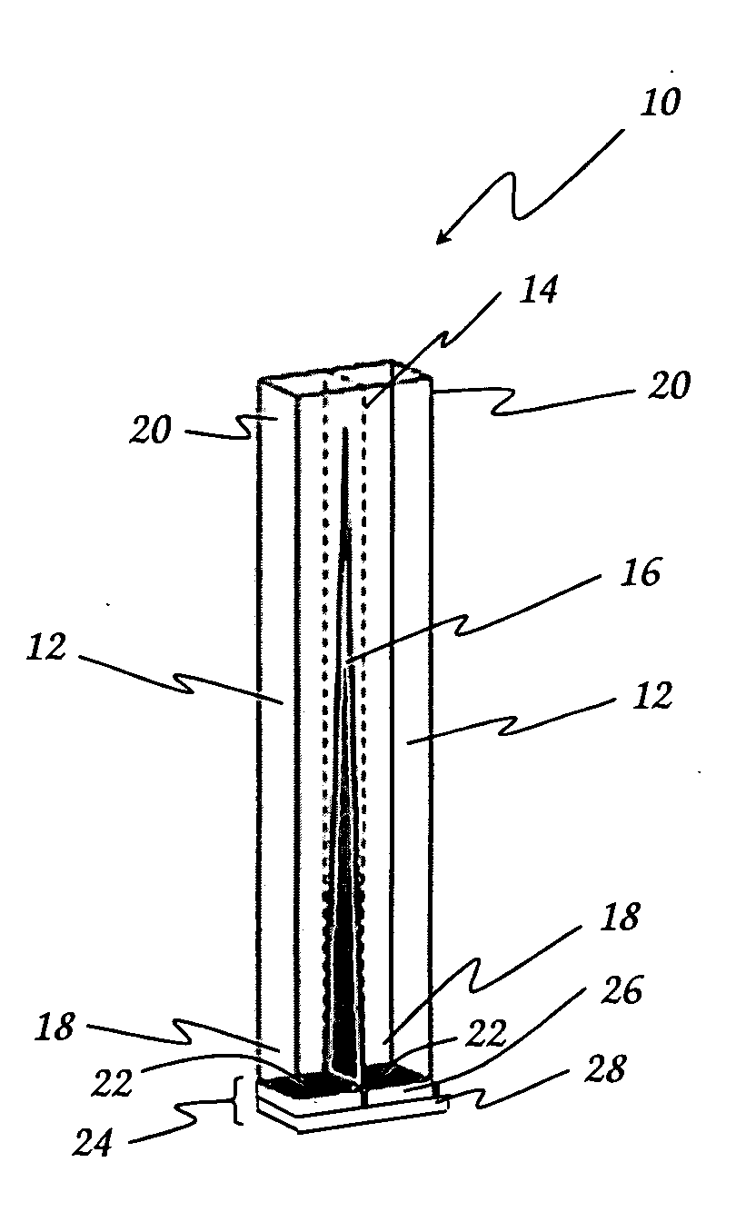
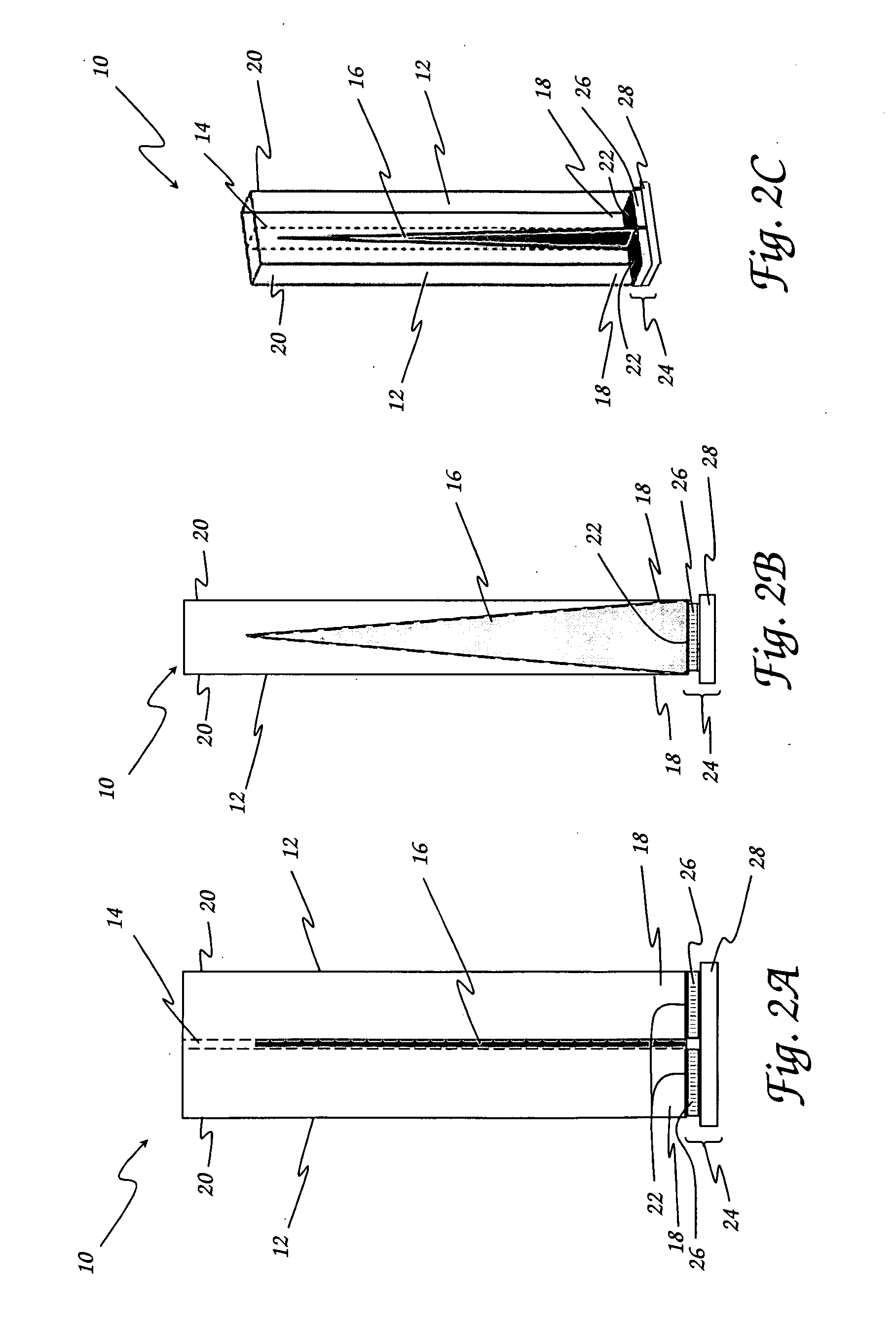
Scintillation detector for positron emission tomography
a scintillation detector and positron emission tomography technology, applied in the field of nuclear medicine imaging devices, can solve the problems of insufficient doi information of conventional pet imaging systems, requirement for additional detector electronics, and limited practical implementation of small cross section crystals and optimization of light sharing between crystals
- Summary
- Abstract
- Description
- Claims
- Application Information
AI Technical Summary
Benefits of technology
Problems solved by technology
Method used
Image

Examples
example
[0019]Zecotek Photonics Inc. has developed three different generations of micro-pixelated avalanche photodiodes (MAPDs), as well as several different variants of their LFS scintillation crystal (Lewellen, T. K., M. Janes, R. S. Miyaoka, and F. Zerrouk, Initial Evaluation of the Scintillator LFS for Positron Emission Tomography Applications, Proceedings of the IEEE Nuclear Science Syposium and Medical Imaging Conference, Rome, (2004): 2915-18). The characteristics of Zecotek's three prototype MAPDs are listed in Table I. Except as noted, the data associated with this Example was taken from MAPD Type 3 devices optically coupled to LFS Type 3 crystals. Zecotek also provided us with some prototype MAPD Type 3 arrays (two 1×2 devices and a 2×2 device), both using 1.1×1.1 mm active cells. Measurements of the current-voltage curves of single devices (or elements in the arrays) were performed. Energy spectra and preliminary timing measurements of single crystal elements were also performed....
PUM
 Abstract
Description
Claims
Application Information